Scan overview
- Thyroid ultrasound uses sound waves to assess the gland’s size, structure and any nodules or cysts, without ionising radiation.
- It may be recommended for a palpable thyroid lump, enlargement or follow-up of a previously identified finding.
- Ultrasound assesses structure rather than hormone production. Blood tests are needed to investigate an overactive or underactive thyroid.
- Most thyroid nodules are benign, but some appearances require specialist assessment or fine-needle aspiration.
- A routine scan is external, usually needs little preparation and does not automatically include a biopsy.
What is a thyroid ultrasound?
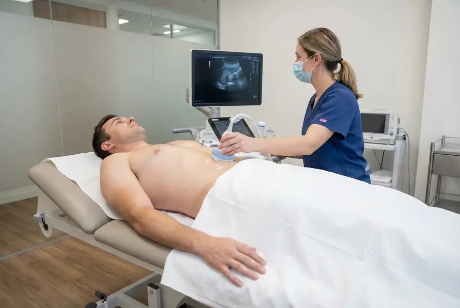
A thyroid ultrasound is a focused examination of the thyroid gland at the front of the lower neck. A trained clinician moves a small handheld probe over the skin to produce real-time images and take measurements.
The thyroid has two lobes joined by a thin bridge of tissue called the isthmus. It produces hormones that influence metabolism and many other body functions. Ultrasound shows the gland’s physical appearance, while thyroid blood tests provide information about hormone production.
The scan can assess overall enlargement, a focal lump or a previously identified nodule. Relevant nearby lymph nodes may also be assessed according to the referral and findings. The examination is tailored to the clinical question rather than being a general check of every neck structure.
Why might you need a thyroid ultrasound?
Your doctor may recommend an ultrasound after examining your neck or reviewing previous results. Reasons can include:
- A lump thought to arise from the thyroid.
- Visible or palpable enlargement of the gland, also called a goitre.
- A thyroid nodule discovered on another imaging examination.
- Monitoring a known thyroid finding where follow-up is clinically indicated.
- Assessment of structural changes in selected thyroid conditions.
- Further evaluation of relevant nearby lymph nodes.
Symptoms such as weight change, tiredness, palpitations or temperature sensitivity may lead to thyroid blood tests, but they do not automatically mean that ultrasound is needed. Your doctor considers whether there is a structural question for imaging to answer.
Persistent voice changes, swallowing difficulty or a growing neck lump need prompt clinical assessment. Breathing difficulty or rapidly worsening neck swelling requires urgent medical attention rather than a routine scan appointment.
Thyroid nodules and cysts
A nodule is a focal area within the thyroid that differs from the surrounding gland. It may be solid, fluid-filled or a mixture of both. Nodules are common, and many are benign; finding a nodule does not automatically mean cancer.
The examiner records relevant measurements and assesses features such as composition, echogenicity, shape, margins and any calcifications. These features are considered together to help guide whether a nodule needs no further action, monitoring or additional assessment.
A simple cyst contains fluid and may have a characteristic appearance. A larger cyst or nodule can sometimes cause pressure symptoms, but the significance depends on its position, size and the wider clinical picture. Not every cyst needs treatment, and drainage is a separate procedure rather than an automatic part of the scan.
Goitre and inflammatory changes
Thyroid enlargement
A goitre means enlargement of the thyroid gland. Ultrasound can help describe its extent and whether the enlargement is diffuse or associated with nodules. There are several possible causes, and the term does not establish hormone activity or a specific diagnosis.
Thyroiditis
Inflammation can alter the gland’s texture or blood-flow pattern. Some appearances may support assessment of thyroiditis, including autoimmune conditions such as Hashimoto’s thyroiditis. The scan is interpreted alongside blood tests and your clinical history.
Graves’ disease and other functional conditions
Ultrasound may provide additional structural or vascular information in selected cases, but an overactive or underactive thyroid is not diagnosed from imaging alone. Hormone levels and, where relevant, antibody tests help establish the cause and guide care.
Can ultrasound identify thyroid cancer?
Ultrasound can identify features that make a nodule more or less concerning, but it cannot reliably determine whether every nodule is benign or malignant. No single feature, including increased blood flow, confirms cancer.
Where a nodule meets the relevant criteria, your doctor may recommend specialist review or ultrasound-guided fine-needle aspiration. This obtains cells for laboratory assessment. Further investigation may still be needed if the sample is inconclusive or does not fully answer the clinical question.
Nearby lymph nodes may be assessed when relevant. An abnormal node can have several causes, so ultrasound findings must be considered with the examination, history and any laboratory results.
Thyroid structure and thyroid function
An ultrasound and a thyroid blood test answer different questions. A gland may have a nodule while producing normal hormone levels, or it may look relatively unremarkable on ultrasound despite abnormal thyroid function.
Blood tests commonly assess thyroid-stimulating hormone and thyroid hormone levels. Further testing is selected according to the results and clinical circumstances. Ultrasound cannot replace these tests or determine treatment requirements by itself.
Your doctor will explain how the imaging findings relate to the symptoms and results discussed during your consultation. Further care may involve your usual doctor, an endocrinologist, an ear, nose and throat specialist or another appropriate clinician.
Types of thyroid ultrasound
Conventional greyscale ultrasound
This is the standard structural examination. It provides images of the gland and nodules in shades of grey, allowing assessment of their appearance and measurements.
Doppler assessment
Colour or power Doppler can add information about blood flow. The colour display is a technical representation of flow, not a direct indication of cancer or whether tissue is healthy. It supports the overall interpretation rather than establishing a diagnosis on its own.
Specialist techniques
Elastography, three-dimensional imaging and contrast-enhanced ultrasound have selected specialist applications. They are not routine requirements for every thyroid nodule and do not replace tissue sampling where it is indicated.
Your team will confirm the examination you need. A standard scan normally requires no injection or sedation; any additional procedure has its own consent and preparation requirements.
What are the limitations of the scan?
Ultrasound cannot reliably confirm or exclude every thyroid cancer, measure hormone production or establish every cause of thyroid inflammation. Some findings require specialist assessment or sampling before a diagnosis can be made.
The view can be limited where tissue extends deep into the chest or lies behind structures that block sound waves. Another imaging technique may be needed to assess the extent of a large goitre or answer a different clinical question.
There is no single normal thyroid measurement that applies to everyone. Size varies with individual circumstances, and the examiner assesses the gland as a whole rather than relying on a single width. Persistent or worsening symptoms should be reviewed even if the report is reassuring.
How to prepare for a thyroid ultrasound
A routine thyroid scan usually requires little preparation. You can generally eat, drink and take your usual medicines unless the centre provides different instructions.
Wear a top with an open or accessible neckline. You may be asked to remove necklaces, scarves or other items that cover the area. Bring relevant previous imaging reports and recent thyroid blood results if available.
Tell the team about previous neck surgery, significant tenderness, difficulty lying flat or extending your neck, or a reaction to ultrasound gel. They can discuss positioning adjustments. If a separate sampling procedure is planned, follow its specific preparation instructions rather than assuming they are the same as for a diagnostic scan.
The procedure: what to expect
- Review and consent: the clinician confirms the referral and any symptoms, explains the examination and answers your questions.
- Positioning: you usually lie on a couch with your neck comfortably exposed. A support may be used to improve the views without forcing your neck backwards.
- Gel application: water-based gel is applied to help sound pass between the probe and skin.
- Scanning: the probe is moved across the front and sides of the lower neck. Sound waves and returning echoes are converted into images on a monitor.
- Images and measurements: the gland and relevant nodules are assessed and recorded, with Doppler or nearby lymph node assessment where useful.
- Completion: the gel is wiped away and the reporting arrangements are explained.
Probe pressure may be uncomfortable over a tender or enlarged area. Tell the examiner if you need them to adjust the technique or pause. Any immediate clinical concern is managed through the centre’s escalation process.
Will I need fine-needle aspiration?
Not every thyroid nodule needs sampling. The decision depends on the ultrasound appearance, size, clinical history and specialist assessment.
Fine-needle aspiration uses a thin needle to collect cells from a selected target, usually with ultrasound guidance. It is different from a routine external examination and requires separate confirmation, consent and arrangements for laboratory analysis.
Your scan price does not automatically include aspiration, a core biopsy or treatment of a cyst. If sampling is recommended, the responsible team will explain its risks, preparation, cost and reporting process. Do not stop blood-thinning medicines or other prescribed treatment unless specifically advised by that team.
Benefits, safety, duration and aftercare
Thyroid ultrasound provides a detailed structural assessment without ionising radiation or an incision. It can document nodules and guide follow-up or sampling decisions when clinically appropriate.
Standard diagnostic ultrasound has no known harmful effects when used appropriately. A skin reaction to the gel is uncommon, and pressure or positioning may cause discomfort if the area is already tender.
The scan commonly takes around 15–30 minutes, although more detailed examinations may take longer. You can usually eat, drink, drive and return to normal activities afterwards. A separate needle procedure has its own aftercare requirements and results timetable.
Getting and understanding your results
Your scan images are reviewed by a consultant radiologist or relevant specialist clinician, who prepares a written imaging report. Results are usually available within three working days of the date of your scan. More complex examinations may require additional specialist review and can take slightly longer.
Once your results are available, a post-scan consultation will be arranged with the same medical doctor who conducted your pre-scan consultation. This continuity allows your doctor to consider the findings alongside the symptoms, medical history and concerns you discussed before your scan.
During the consultation, your doctor will explain the report in detail, answer your questions and discuss what the findings mean for you. Where further assessment or care is needed, they will explain the recommended next steps, which may include additional investigations, a specialist referral or follow-up with your usual healthcare team.
How much does a private thyroid ultrasound cost?
Private ultrasound scans with PrivateScans start from £195. This is our general ultrasound starting price. Your confirmed cost depends on the centre, scan coverage and any specialist requirements.
A routine thyroid examination is different from a wider neck assessment or a sampling procedure. Your consultation helps establish the appropriate investigation, and the examination and price will be confirmed before you proceed.
Book a thyroid ultrasound
Your booking includes a 30-minute private telephone consultation with a medical doctor at a convenient time. You can discuss the lump or symptoms, thyroid blood results, relevant medical history and previous investigations so your doctor can advise on the appropriate next step.
Where ultrasound is clinically appropriate, a written referral is prepared and your scan can be arranged at a suitable centre. Your report is then discussed with the same doctor, who will explain the findings and any necessary next steps. Explore available ultrasound appointments or return to our ultrasound body parts directory. You can also read our neck ultrasound guide for broader neck concerns.
FAQs
Can ultrasound diagnose an underactive or overactive thyroid?
Not by itself. Ultrasound assesses the gland’s structure, while blood tests assess hormone production. Your doctor interprets these results alongside your symptoms and medical history.
Does a thyroid nodule mean cancer?
No. Many nodules are benign. The scan assesses a combination of features to help determine whether monitoring, sampling or specialist review is needed.
What does thyroid cancer look like on ultrasound?
Some features can raise concern, but there is no single appearance that reliably confirms cancer. Blood-flow changes alone are not diagnostic. Tissue sampling or specialist assessment may be needed.
Will I need a biopsy?
Not every nodule requires sampling. The decision depends on the ultrasound findings and clinical circumstances. A routine diagnostic scan does not automatically include fine-needle aspiration or another biopsy.
Is there a single normal thyroid size?
No. Thyroid size varies between individuals. The examiner records relevant measurements and considers the overall appearance rather than relying on one universal width.
Can I take my thyroid medication before the scan?
You can usually take your prescribed medicines as normal for a routine ultrasound. Follow any specific instructions from your centre and do not stop treatment without advice.
Will the scan be painful?
It is an external examination. Probe pressure or neck positioning may be uncomfortable if the area is tender. Tell the examiner so they can adjust the technique or provide support.
What’s included with your scan?
What is included with your scan:
- A pre-scan telephone consultation with a member of our medical team.
- A written medical referral prepared from the information discussed during your consultation, where clinically appropriate.
- Your scan at the chosen scanning centre.
- A written imaging report prepared by a consultant radiologist or relevant specialist clinician.
- Access to downloadable copies of your scan images where required.
- A post-scan consultation to discuss your results and next steps where needed.