Scan overview
- Liver ultrasound uses sound waves to assess the liver’s size, shape and internal appearance without ionising radiation.
- It may be recommended after abnormal liver blood tests, for persistent upper abdominal symptoms or to monitor a known condition.
- The examination can identify changes associated with fatty liver, some liver lesions and features that may suggest advanced chronic liver disease.
- Doppler ultrasound and elastography provide different information and are not automatically included in a routine liver scan.
- Findings are considered alongside your medical history and blood tests. Further imaging or specialist assessment may be needed to establish a diagnosis.
What is a liver ultrasound scan?
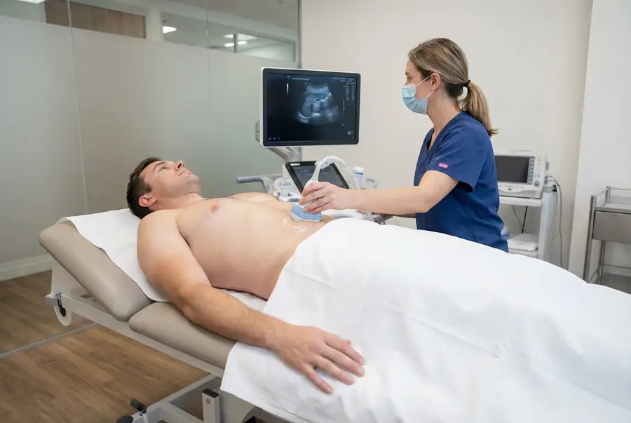
A liver ultrasound, also called hepatic ultrasound or hepatic sonography, is an external examination of the liver and relevant nearby structures. A trained clinician moves a small handheld probe across the upper abdomen, usually on the right-hand side beneath the ribs.
The probe sends high-frequency sound waves into the body and receives the returning echoes. A computer converts these signals into real-time images, allowing the examiner to assess the liver’s outline, tissue appearance and visible abnormalities.
The liver performs many essential functions, including processing nutrients, producing bile and helping remove substances from the blood. Ultrasound provides information about its structure, but it does not directly measure all aspects of liver function. Blood tests and a clinical assessment remain important parts of understanding your liver health.
Why might you need a liver ultrasound?
Your doctor may recommend an ultrasound to investigate a possible liver or biliary problem, assess an unexpected blood test result or monitor a condition that has already been diagnosed. Reasons for imaging can include:
- Abnormal liver blood tests, including raised liver enzymes.
- Persistent discomfort in the upper right abdomen.
- Jaundice, where the skin or whites of the eyes appear yellow.
- Symptoms such as dark urine, pale stools or persistent itching, where a liver or bile duct cause is suspected.
- Assessment of suspected fatty liver in the context of metabolic risk factors or alcohol use.
- Follow-up of a previously identified liver lesion or chronic liver condition.
- A specialist surveillance programme for people with particular established risk factors.
These symptoms can have several causes, and not everyone needs an ultrasound. Your doctor will consider the pattern of symptoms, medicines, alcohol intake, medical history and blood test results before advising on the appropriate investigation.
New jaundice needs prompt medical assessment. Severe abdominal pain, particularly with fever, vomiting or feeling very unwell, should be assessed urgently rather than waiting for a routine scan.
What does a liver ultrasound show?
The examiner assesses the liver’s size, contour and internal texture, looking for general changes throughout the organ and any focal abnormality. They may record measurements and assess relevant blood vessels and bile ducts, according to the requested examination.
Ultrasound can provide information about:
- Liver size and shape: enlargement or an altered contour can help guide further investigation.
- Tissue appearance: changes in brightness or texture may suggest fat accumulation or chronic liver disease, although these appearances are not always specific.
- Focal lesions: cysts, some benign growths and suspicious masses may be visible.
- Bile ducts: widening of visible ducts can suggest an obstruction that needs further assessment.
- Related findings: fluid in the abdomen or changes affecting nearby structures may be identified within the examination’s coverage.
A liver-focused scan may be arranged as part of an upper abdominal ultrasound. Whether the gallbladder, pancreas, spleen or other organs are included depends on the referral and appointment. Your team will confirm the scope before you proceed.
Fatty liver and chronic liver disease
Fat accumulation in the liver
A liver containing excess fat may appear brighter on ultrasound than expected. A report may describe this as increased echogenicity or an appearance consistent with steatosis. Your doctor will explain this alongside blood tests and relevant factors such as metabolic health, alcohol use and medication.
A routine scan cannot reliably identify every degree of fat accumulation, distinguish all causes of fatty liver or determine whether inflammation and scarring are present. Further assessment may be appropriate even if the ultrasound findings are mild.
Fibrosis and cirrhosis
More advanced chronic liver disease may alter the liver’s surface and texture. Ultrasound can show a nodular contour, changes in liver shape or findings associated with portal hypertension, such as fluid in the abdomen. The liver is not necessarily small in every person with cirrhosis.
Early fibrosis may not be visible on routine ultrasound, and a normal-looking liver does not exclude chronic liver disease. Blood-based assessment, elastography and specialist review may be needed to evaluate the likelihood and extent of scarring.
Hepatitis
Hepatitis means inflammation of the liver. Ultrasound findings can be non-specific or normal, so the scan cannot establish or exclude hepatitis on its own. The cause and clinical significance are usually assessed with your history, blood tests and other investigations where appropriate.
Liver cysts, masses and possible cancer
Ultrasound may identify a focal lesion: an area that looks different from the surrounding liver. Some lesions, such as a simple fluid-filled cyst, have characteristic appearances. Others need closer assessment before their nature can be established.
Haemangiomas are benign growths made up of blood vessels and can sometimes have a typical ultrasound appearance. However, a solid lesion cannot always be classified confidently from routine ultrasound alone. Its appearance, size and your clinical history influence whether further investigation is recommended.
Ultrasound can detect some liver cancers, but it does not detect every tumour, particularly small lesions or abnormalities in a liver that is difficult to visualise. It cannot reliably confirm or exclude cancer by itself. CT, MRI, specialist contrast-enhanced ultrasound or, in selected cases, a biopsy may be advised.
Regular liver cancer surveillance is offered to particular higher-risk groups under clinical guidance. It is not a universal requirement for everyone with diabetes, fatty liver or a history of alcohol use. Your doctor can explain whether a specialist surveillance programme is appropriate for you.
Bile ducts, gallbladder and blood vessels
Where included in the examination, ultrasound can assess the gallbladder for stones and features that may suggest inflammation. It can also show widening of the bile ducts, which may indicate impaired bile drainage. Not every bile duct stone or underlying cause of obstruction is visible, so further imaging may be required.
Doppler assessment can examine the direction and pattern of blood flow in relevant liver vessels, including the portal vein and hepatic veins. It may support investigation of suspected vascular abnormalities, a clot or changes associated with portal hypertension.
These findings require clinical interpretation. Ultrasound does not provide a complete assessment of every vascular or biliary condition. MRI, MRCP or CT may be more appropriate when the clinical question needs more detailed imaging.
Routine ultrasound, Doppler and elastography
Standard liver ultrasound
Conventional ultrasound produces images of the liver and relevant surrounding structures. It is commonly used as an initial structural assessment.
Doppler ultrasound
Doppler techniques add information about blood flow. They may be used during a routine examination when useful, or requested as a more focused assessment of the liver’s circulation.
Liver elastography
Elastography estimates liver stiffness and can help assess the likelihood of fibrosis. Different techniques are available, including transient elastography and shear-wave elastography. Stiffness is influenced by factors other than scarring, so the measurement needs to be interpreted in context.
Elastography is not automatically included in a standard liver ultrasound. If it is clinically indicated, the team will confirm the appropriate examination, preparation, availability and price.
What are the limitations of liver ultrasound?
Image quality can be affected by bowel gas, the position of the liver, body habitus and your ability to maintain the requested position or breathing pattern. Sometimes parts of the organ cannot be fully assessed, and the report may explain these limitations.
A normal scan does not rule out every liver condition, early fibrosis or a small tumour. An abnormal finding also does not automatically mean cancer or severe disease. Some changes are incidental and benign, while others require additional tests to clarify their significance.
Your doctor may recommend blood tests, a liver MRI, a wider CT examination or specialist assessment, depending on the clinical question. The most useful next test depends on the findings and your individual circumstances.
How to prepare for a liver ultrasound
Your centre will provide preparation instructions for the examination you have booked. Fasting is often requested before a liver or upper abdominal ultrasound to improve the assessment of relevant structures, particularly the gallbladder, and reduce interference from bowel contents.
The length of fasting and the advice about water vary between examinations and centres. Follow your appointment instructions rather than a general rule. Do not assume that you need to avoid all fluids. If the instructions are unclear, contact the team before your appointment.
Tell the team if you have diabetes, a condition affected by fasting or medicines that need to be taken with food. They can advise on suitable arrangements. Do not stop prescribed medicines unless you have been instructed to do so.
Wear comfortable clothing that allows access to your upper abdomen. Bring relevant previous reports, recent liver blood test results if available and details of your medicines. Let the team know about mobility needs or a previous reaction to ultrasound gel.
The procedure: what to expect
- Review and explanation: the examiner confirms the reason for your scan, checks preparation and explains the examination before obtaining your consent.
- Positioning: you usually lie on your back on an examination couch with your upper abdomen exposed. You may be asked to turn slightly onto your side or raise an arm.
- Gel application: water-based gel is placed on the skin to help transmit the sound waves.
- Scanning: the probe is moved across the upper abdomen and around the rib margin. Gentle pressure is applied to obtain views from different angles.
- Breathing and measurements: you may be asked to take a breath and hold it briefly. Representative images and relevant measurements are recorded, with additional techniques where requested.
- Completion: the gel is removed and the team explains the reporting arrangements and any immediate instructions.
You cannot normally hear or feel the ultrasound waves. Probe pressure may be uncomfortable over a tender area; tell the examiner if you need them to adjust the pressure or positioning.
Safety, duration and aftercare
A routine liver ultrasound uses no ionising radiation and normally requires no injection or sedation. Standard diagnostic ultrasound has no known harmful effects when used appropriately. A skin reaction to the gel is uncommon.
The examination often takes around 15–30 minutes, although a more detailed assessment or additional Doppler imaging may take longer. Allow time for registration and any preparation required by the centre.
You can usually eat, drink, drive and return to normal activities afterwards, unless you have been given other instructions. A separate procedure, such as a biopsy, has different preparation and aftercare requirements and is not part of a routine liver ultrasound.
Getting and understanding your results
Your scan images are reviewed by a consultant radiologist or relevant specialist clinician, who prepares a written imaging report. Results are usually available within three working days of the date of your scan. More complex examinations may require additional specialist review and can take slightly longer.
Once your results are available, a post-scan consultation will be arranged with the same medical doctor who conducted your pre-scan consultation. This continuity allows your doctor to consider the findings alongside the symptoms, medical history and concerns you discussed before your scan.
During the consultation, your doctor will explain the report in detail, answer your questions and discuss what the findings mean for you. Where further assessment or care is needed, they will explain the recommended next steps, which may include additional investigations, a specialist referral or follow-up with your usual healthcare team.
How much does a private liver ultrasound cost?
Private ultrasound scans with PrivateScans start from £195. This is our general ultrasound starting price. The confirmed cost depends on the centre, the scope of the examination and any specialist techniques required.
A routine liver examination, a wider abdominal scan and a dedicated elastography assessment are not necessarily the same service. Your medical consultation helps establish the appropriate investigation, and the examination and its price will be confirmed before you proceed.
Book a liver ultrasound
Your booking includes a 30-minute private telephone consultation with a medical doctor at a convenient time. You can discuss your symptoms, liver blood test results, relevant medical history and any previous investigations so your doctor can advise whether ultrasound is an appropriate next step.
Where imaging is clinically appropriate, a referral is prepared and your scan can be arranged at a suitable centre. Your results are then discussed with the same doctor, with an explanation of any recommended next steps. Explore available ultrasound appointments or visit our ultrasound body parts directory.
FAQs
Can I drink water before my liver ultrasound?
Follow the instructions provided by your scanning centre. Fasting is often requested, but small amounts of water may be permitted depending on the examination. Contact the team if you are unsure, rather than avoiding all fluids without advice.
Can ultrasound show fatty liver?
Ultrasound can show changes consistent with fat accumulation, often described as increased brightness or echogenicity. It may miss mild changes and cannot reliably determine whether inflammation or fibrosis is present. Your doctor will interpret the findings with blood tests and your medical history.
Can a normal scan rule out liver disease?
No. Some liver conditions, early fibrosis and small lesions may not be visible. If symptoms or blood tests remain concerning, your doctor may recommend further assessment even when the scan is reported as normal.
Can liver ultrasound detect cancer?
It can identify some suspicious lesions but cannot detect every cancer or reliably confirm the nature of every mass. Further imaging or specialist assessment may be necessary. A biopsy is used in selected cases rather than being required for every liver lesion.
Is elastography included in my scan?
Not automatically. Elastography measures liver stiffness and is a separate technique from routine structural ultrasound. Your doctor can advise whether it is appropriate, and the team will confirm availability, preparation and cost before booking.
Does an abnormal liver ultrasound mean something serious?
Not necessarily. Findings such as simple cysts or fatty changes are common, and their significance depends on the full clinical picture. Your follow-up consultation explains what the report means and whether monitoring, further tests or specialist care are needed.
Will I receive my results at the appointment?
Some immediate observations may be discussed where appropriate, but your formal report and clinical interpretation are provided through the reporting and follow-up process. Results are usually available within three working days of your scan; more complex examinations may take longer.
What’s included with your scan?
What is included with your scan:
- A pre-scan telephone consultation with a member of our medical team.
- A written medical referral prepared from the information discussed during your consultation, where clinically appropriate.
- Your scan at the chosen scanning centre.
- A written imaging report prepared by a consultant radiologist or relevant specialist clinician.
- Access to downloadable copies of your scan images where required.
- A post-scan consultation to discuss your results and next steps where needed.