Scan overview
- Prostate ultrasound uses sound waves to assess the gland’s size and selected structural features without ionising radiation.
- The examination may be performed externally over the lower abdomen or with a transrectal probe, depending on the clinical question and the service arranged.
- Ultrasound alone cannot reliably diagnose or exclude prostate cancer. PSA testing, clinical assessment, MRI and sometimes biopsy have different roles.
- A biopsy is a separate invasive procedure and is not automatically included in a diagnostic ultrasound appointment.
- Your medical consultation helps establish the appropriate examination, with preparation, consent and price confirmed before you proceed.
What is a prostate ultrasound?
A prostate ultrasound produces real-time images of the prostate gland using a handheld or specialised internal probe. It can provide measurements of gland size and information about selected structural changes.
The prostate sits below the bladder, surrounds the upper part of the urethra and lies in front of the rectum. It produces fluid that contributes to semen. Enlargement or other prostate problems may affect urination, although urinary symptoms can also arise from the bladder or other causes.
Because the gland is close to the rectum, a transrectal ultrasound can obtain detailed local views. An external transabdominal examination can also estimate prostate size in suitable circumstances, often alongside bladder assessment. The most appropriate approach depends on the information required and your individual needs.
Why might you need prostate imaging?
Your doctor may consider prostate assessment when investigating symptoms such as:
- Difficulty starting urination or a weak stream.
- Urinary frequency, urgency or waking at night to pass urine.
- A feeling that the bladder has not emptied fully.
- Persistent pelvic or perineal discomfort.
- Blood in the urine or semen, following appropriate clinical review.
- A known prostate finding that needs further assessment or monitoring.
These symptoms do not establish a particular diagnosis or automatically mean that ultrasound is the best next test. Your doctor may recommend urine tests, PSA testing, an examination or referral to a urologist.
An abnormal PSA result or concerning examination may require a prostate cancer investigation pathway rather than a standalone ultrasound. Sudden inability to pass urine, or urinary symptoms with fever and feeling acutely unwell, requires urgent medical assessment. Blood in the urine also needs prompt review.
What can a prostate ultrasound show?
Prostate size and enlargement
Ultrasound can estimate prostate volume and help assess enlargement. Benign prostatic enlargement is a common non-cancerous change, but size alone does not show how severe symptoms are or establish whether there is bladder outlet obstruction.
Cysts and calcifications
The scan may identify fluid-filled cysts or small calcifications within the gland. These can be incidental, and their significance depends on the appearance, location and clinical context. A cyst is not necessarily an abscess or a collection of pus.
Selected inflammatory or structural abnormalities
Ultrasound may contribute to assessment of a suspected collection or abscess in a specialist setting. Prostatitis is primarily a clinical diagnosis supported by appropriate tests; a normal scan does not exclude it.
Related structures
A specifically requested examination may assess the seminal vesicles or selected findings relevant to ejaculatory duct obstruction. A combined external scan may also assess the bladder and residual urine. These elements are not automatically included in every prostate appointment.
Prostate ultrasound, PSA and possible cancer
PSA is a protein produced by the prostate and measured in a blood test. A raised result can occur in several circumstances and does not, by itself, confirm cancer. Equally, ultrasound cannot provide reassurance that cancer is absent solely because the gland appears unremarkable.
Some cancers are not visible on conventional ultrasound, while benign changes can also look abnormal. A description of a lighter or darker area is not a reliable diagnosis on its own.
Where prostate cancer is suspected, your doctor may recommend specialist assessment, prostate MRI and, where appropriate, biopsy. Ultrasound is often used to guide sampling or measure the gland, but it does not replace the wider diagnostic pathway.
Your consultation will consider why imaging is being requested so that the examination is appropriate for the clinical question, rather than using a routine ultrasound as a standalone cancer screening test.
Types of prostate ultrasound
Transabdominal ultrasound
A probe is moved over the lower abdomen, usually with a full bladder. This external approach can estimate prostate size and assess the bladder where included. It generally provides less detailed local views of the prostate than a transrectal examination.
Transrectal ultrasound (TRUS)
A slim, covered and lubricated probe is gently placed in the rectum with your consent. Its proximity to the prostate allows measurements and detailed local imaging. It may be used for a diagnostic question or as part of a separately arranged procedure.
Ultrasound guidance for transperineal biopsy
In a transperineal biopsy, sampling needles pass through the skin between the scrotum and anus. An ultrasound probe, often positioned in the rectum, helps guide the needles. The needle route and the imaging approach are different parts of the procedure.
A biopsy is not simply another name for a prostate ultrasound. Its availability, indication, anaesthetic, preparation and aftercare must be confirmed separately.
Consent, privacy and comfort
Before an internal examination, the clinician explains its purpose, what it involves and the alternatives available. You can ask questions, decline the examination or ask for it to stop at any point.
A chaperone is offered for an intimate examination in accordance with the centre’s arrangements. Privacy is maintained while you change and during the examination, with appropriate covering.
Tell the team about previous rectal surgery, significant rectal pain, bleeding, allergies or concerns about an internal examination. A transrectal approach may be unsuitable after certain operations or in other circumstances. Your doctor can consider an external examination, MRI or another appropriate investigation.
What are the limitations of prostate ultrasound?
A standard ultrasound cannot reliably exclude prostate cancer, establish every cause of urinary symptoms or diagnose all forms of prostatitis. It does not assess erectile function or explain every cause of pelvic pain.
Image quality depends on the approach and available scanning window. Bowel contents, anatomy or difficulty tolerating the examination may affect the views. An external scan also relies on suitable bladder filling.
Bladder emptying measurements and prostate size may provide useful clues, but they do not replace other functional tests where needed. Your doctor may recommend a urinary tract ultrasound, additional laboratory tests, MRI or urological assessment according to the clinical concern.
How to prepare for a prostate ultrasound
Follow the instructions for the examination you have booked. For an external scan, the centre may ask you to drink water and avoid passing urine before the initial images. Contact the team if you have a fluid restriction, a catheter or difficulty holding urine.
Preparation for TRUS varies. Do not use an enema, take antibiotics or change your medicines unless the team specifically instructs you to do so. Routine diagnostic ultrasound and a biopsy have different preparation requirements.
Bring relevant previous reports, PSA results if available and a list of medicines. Tell the centre about blood-thinning treatment, allergies, rectal conditions or surgery, and any mobility or positioning needs.
Do not stop prescribed medicines, including anticoagulants or antiplatelets, without advice from the team responsible for your care. If fasting or other special preparation is necessary, this will be explained before the appointment.
The procedure: what to expect
External examination
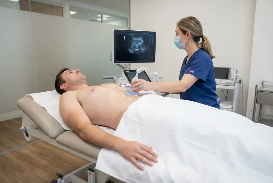
The clinician confirms your symptoms, explains the scan and obtains consent. You usually lie on your back with the lower abdomen exposed. Gel is applied and the probe is moved over the skin to obtain images and measurements. Bladder assessment may be performed, including a post-void measurement where requested.
Transrectal examination
After discussion and consent, you are usually positioned on your side with your knees bent. A cleaned and disinfected probe is fitted with a single-use protective cover and lubricating gel, then gently placed in the rectum. The clinician adjusts it to obtain the required views and measurements.
Pressure or discomfort can occur. Tell the examiner if you are in pain so they can pause or adapt the procedure. The probe is removed once imaging is complete, and you are given privacy to clean away gel and dress.
Sampling does not take place automatically. If a biopsy has been separately arranged, the responsible team will explain and obtain consent for that procedure. Any immediate concern is managed through the centre’s clinical escalation process.
If a prostate biopsy is recommended
A biopsy obtains small samples of prostate tissue for laboratory analysis. It requires a separate decision based on the wider assessment, including the clinical findings and imaging where relevant.
The route of sampling, anaesthetic and preparation depend on the procedure arranged. The team will explain risks such as bleeding, infection and difficulty passing urine, along with instructions about medicines and recovery.
Biopsy findings follow a different reporting process from a routine ultrasound report and may take longer. Your specialist team will explain when to expect the pathology results and how they will be discussed. Seek advice promptly if you develop concerning symptoms after any procedure, following the discharge instructions provided.
Safety, duration and aftercare
Standard diagnostic ultrasound uses no ionising radiation and has no known harmful effects when used appropriately. An external scan is non-invasive; TRUS involves an internal probe and can cause pressure or discomfort.
Tell the team about a latex or gel sensitivity so suitable materials can be selected. A diagnostic examination commonly takes around 15–30 minutes, with extra time for discussion, changing and any preparation.
You can usually return to normal activities after diagnostic imaging unless advised otherwise. If sedation, anaesthetic or a biopsy is involved, driving and activity instructions may differ. Follow the advice specific to the procedure you have had.
Getting and understanding your results
Your scan images are reviewed by a consultant radiologist or relevant specialist clinician, who prepares a written imaging report. Results are usually available within three working days of the date of your scan. More complex examinations may require additional specialist review and can take slightly longer.
Once your results are available, a post-scan consultation will be arranged with the same medical doctor who conducted your pre-scan consultation. This continuity allows your doctor to consider the findings alongside the symptoms, medical history and concerns you discussed before your scan.
During the consultation, your doctor will explain the report in detail, answer your questions and discuss what the findings mean for you. Where further assessment or care is needed, they will explain the recommended next steps, which may include additional investigations, a specialist referral or follow-up with your usual healthcare team.
How much does a private prostate ultrasound cost?
Private ultrasound scans with PrivateScans start from £195. This is our general ultrasound starting price and does not guarantee the price or availability of a specialist prostate examination.
The confirmed cost depends on the centre, the approach and scan coverage. TRUS, specialist assessment and biopsy may have separate arrangements and prices. Your consultation helps establish the appropriate service, and the examination and cost will be confirmed before you proceed.
Book a prostate ultrasound
Your booking includes a 30-minute private telephone consultation with a medical doctor at a convenient time. You can discuss urinary symptoms, PSA results, relevant medical history and previous investigations so your doctor can advise on an appropriate next step.
Where ultrasound is clinically appropriate, a written referral is prepared and the team can help arrange a suitable examination, subject to the service available at the centre. Your report is discussed with the same doctor, who will explain the findings and any necessary next steps. Explore available ultrasound appointments or visit our ultrasound body parts directory.
FAQs
Is prostate ultrasound always an internal scan?
No. An external transabdominal examination can assess selected questions, including prostate size and bladder findings. TRUS uses a probe in the rectum for more detailed local views. The approach depends on the referral and the service arranged.
Can ultrasound rule out prostate cancer?
No. Some cancers are not visible on conventional ultrasound, and benign changes may also look abnormal. Clinical assessment, PSA, MRI and sometimes biopsy have different roles in investigating possible cancer.
Does a raised PSA mean I need an ultrasound?
Not necessarily. A raised result needs clinical interpretation, and your doctor may recommend specialist assessment or MRI rather than a standalone ultrasound. The appropriate pathway depends on your circumstances.
Will a biopsy be done during the scan?
Not automatically. A biopsy is a separate invasive procedure requiring specific booking, preparation and consent. It should not be assumed to be included in a diagnostic scan.
Is TRUS painful?
It can cause pressure or discomfort. Tell the clinician if you are in pain; you can ask them to pause or stop. The examination should be explained beforehand, with privacy and chaperone arrangements discussed.
Should I stop blood-thinning medication?
Do not stop prescribed medicines without advice. Diagnostic ultrasound and biopsy have different requirements. The team responsible for any procedure will give individual instructions where a change is necessary.
Do I need a full bladder?
Often for an external prostate and bladder examination, but instructions vary by approach. Follow the guidance for your appointment and tell the team about fluid restrictions or difficulty holding urine.
What’s included with your scan?
What is included with your scan:
- A pre-scan telephone consultation with a member of our medical team.
- A written medical referral prepared from the information discussed during your consultation, where clinically appropriate.
- Your scan at the chosen scanning centre.
- A written imaging report prepared by a consultant radiologist or relevant specialist clinician.
- Access to downloadable copies of your scan images where required.
- A post-scan consultation to discuss your results and next steps where needed.