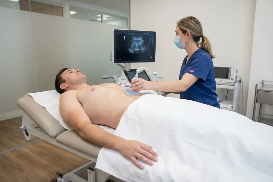
Scan overview
- Testicular ultrasound, also called scrotal ultrasound, uses sound waves to assess the testicles and surrounding scrotal structures without ionising radiation.
- It can investigate a lump, swelling or persistent discomfort and help establish whether a finding lies within or outside a testicle.
- Doppler imaging may assess blood flow when relevant to the clinical question.
- The scan is performed externally over the scrotal skin, with privacy, explanation and consent throughout.
- Sudden severe testicular pain needs emergency assessment; do not wait for a routine private scan.
What is a testicular ultrasound?
A testicular ultrasound is a targeted examination of the testicles and relevant surrounding tissues in the scrotum. A trained clinician gently moves a small handheld probe over the skin to obtain real-time images.
The testicles produce sperm and hormones, including testosterone. Nearby structures include the epididymis, where sperm mature, and the spermatic cord. Ultrasound can help identify which structure accounts for a palpable lump or swelling.
The examination commonly assesses both sides for comparison, even if symptoms are mainly on one side. It is not a prostate scan or a complete investigation of fertility or hormone production. The referral and clinical history guide the examination.
Why might you need a testicular ultrasound?
Your doctor may recommend the scan to assess:
- A new lump or a change in the size or feel of a testicle.
- Persistent or recurrent scrotal discomfort after clinical assessment.
- Swelling or an apparent fluid collection.
- A suspected epididymal cyst or varicocele.
- Selected findings following injury or inflammation.
- A known abnormality that needs further assessment or monitoring.
A new testicular lump should be reviewed promptly, even when it is painless. Many scrotal findings are benign, but their location and appearance need assessment rather than assumptions.
Sudden severe testicular pain, especially with nausea, vomiting or rapid swelling, requires emergency medical assessment. Testicular torsion is time-sensitive, and arranging a routine scan must not delay urgent care.
What does the scan show?
The examiner assesses the size, shape and internal appearance of the testicles, the epididymides and relevant surrounding tissues. The scan may establish whether a finding is within the testicle or in an adjacent structure.
Useful observations can include:
- Whether a palpable abnormality is solid, fluid-filled or mixed.
- Fluid around a testicle or a cyst in an adjacent structure.
- Enlarged veins or selected inflammatory changes.
- A focal lesion that requires further clinical assessment.
- Blood-flow patterns when Doppler imaging is appropriate.
These findings are interpreted with the symptoms and examination. An ultrasound appearance alone does not answer every question about the cause of pain or the nature of a lesion.
Conditions that may be assessed
Cysts and hydroceles
Epididymal cysts are fluid-filled findings outside the testicle and can have a characteristic benign appearance. A hydrocele is a collection of fluid around the testicle that can cause swelling. The significance and need for treatment depend on your symptoms and the full assessment.
Varicoceles
A varicocele is an enlargement of veins in the scrotum. The examiner may assess the veins in different positions and ask you to perform a brief bearing-down manoeuvre. Its relevance to discomfort or fertility is considered clinically rather than inferred from its presence alone.
Inflammatory changes
Ultrasound may show changes associated with inflammation of the epididymis or testicle. Symptoms, examination and urine or other tests may be needed to establish the cause and guide treatment.
Other structural findings
Selected abnormalities following injury, a suspected hernia or the position of a testicle may require tailored assessment. Ultrasound cannot locate every undescended testicle or replace specialist review where this is needed.
Testicular lumps and possible cancer
Ultrasound is useful for assessing a suspected testicular mass and distinguishing many intratesticular findings from lesions in surrounding tissues. A solid lesion within a testicle may require prompt urological assessment.
A cystic appearance can support a benign explanation in the right context, but not every lump can be confidently classified from imaging alone. Ultrasound cannot determine every tumour type or replace specialist evaluation.
Your doctor will explain any recommended next steps, which may include specialist review, blood tests or additional imaging. Do not delay assessment of a new or changing lump while waiting to see whether it becomes painful.
Doppler imaging and urgent symptoms
Doppler techniques provide information about blood flow within the testicles and nearby vessels. They may help assess inflammatory changes, enlarged veins or a concern about blood supply.
Testicular torsion occurs when twisting compromises the testicle’s blood supply. It is an emergency clinical problem. An outpatient ultrasound is not an appropriate substitute for urgent assessment, and a reassuring scan does not necessarily settle every concern about intermittent or early torsion.
If you develop sudden severe pain or rapidly worsening symptoms, seek emergency medical attention rather than waiting for your booked appointment or routine results.
What are the limitations of the scan?
Ultrasound may not explain every cause of scrotal pain and cannot directly measure sperm quality or hormone production. Fertility and endocrine concerns may need separate clinical assessment and laboratory tests.
Appearance can overlap between different conditions. Some findings need specialist review, repeat imaging or further investigations to clarify their significance.
A normal report does not mean persistent or changing symptoms should be ignored. Discuss continuing concerns with your doctor, and seek urgent care for acute symptoms regardless of a previous scan result.
Privacy, consent and comfort
The clinician explains the examination and obtains your consent before proceeding. You can ask questions, request a pause or ask for the scan to stop.
You are given privacy to change and appropriate covering. A chaperone is offered for an intimate examination in accordance with the centre’s arrangements. You can discuss chaperone arrangements or an examiner preference with the centre beforehand, subject to availability.
Tell the team if you feel anxious, have significant tenderness or need assistance with positioning. The examination is performed externally, and pressure can be adjusted to your comfort and the views needed.
How to prepare for a testicular ultrasound
A routine testicular ultrasound usually requires no special preparation. You can generally eat, drink and take normal medicines unless the centre advises otherwise. A full bladder is not usually required for a scrotal-only examination.
Wear comfortable clothing that is easy to adjust. There is no need to shave or groom before the scan. Bring relevant previous reports and tell the team about the exact location of a lump, any injury or operation, and how the symptoms have changed.
Let the centre know about a wound, dressing, mobility needs or a previous reaction to ultrasound gel. Follow any different instructions if another examination is planned at the same appointment.
The procedure: what to expect
- Discussion and consent: the examiner confirms your symptoms and referral, explains the scan and answers your questions.
- Positioning: you usually lie on your back with the relevant area exposed and the rest of your body covered. A towel or other support may be used to position the scrotum comfortably.
- Gel application: water-based gel is applied to the skin to improve transmission of the sound waves.
- Scanning: the probe is moved gently over the scrotum. High-frequency sound waves and returning echoes are converted into images on a monitor.
- Images and measurements: both sides and relevant findings are recorded, with Doppler assessment or a position change where useful.
- Completion: you are given tissue and privacy to remove gel and dress. The team explains the reporting arrangements.
Tell the examiner if probe pressure is painful. Any immediate clinical concern is managed through the centre’s escalation process rather than simply waiting for a routine report.
Safety, duration and aftercare
Standard diagnostic ultrasound has no known harmful effects when used appropriately and does not use ionising radiation. A routine scrotal scan normally requires no injection or sedation.
Probe pressure may cause discomfort if the area is tender, and a reaction to the gel is uncommon. Let the examiner know about pain or sensitivities.
The examination often takes around 15–30 minutes, although duration varies with the findings and assessment required. You can usually drive and return to normal activities afterwards unless advised otherwise. Continue to follow any clinical advice relating to your symptoms.
Getting and understanding your results
Your scan images are reviewed by a consultant radiologist or relevant specialist clinician, who prepares a written imaging report. Results are usually available within three working days of the date of your scan. More complex examinations may require additional specialist review and can take slightly longer.
Once your results are available, a post-scan consultation will be arranged with the same medical doctor who conducted your pre-scan consultation. This continuity allows your doctor to consider the findings alongside the symptoms, medical history and concerns you discussed before your scan.
During the consultation, your doctor will explain the report in detail, answer your questions and discuss what the findings mean for you. Where further assessment or care is needed, they will explain the recommended next steps, which may include additional investigations, a specialist referral or follow-up with your usual healthcare team.
How much does a private testicular ultrasound cost?
Private ultrasound scans with PrivateScans start from £195. This is our general ultrasound starting price. Your confirmed cost depends on the scanning centre, examination coverage and any specialist requirements.
Your medical consultation helps establish the appropriate investigation. The examination and price will be confirmed before you proceed. A diagnostic scan does not automatically include specialist treatment or a separate fertility assessment.
Book a testicular ultrasound
Your booking includes a 30-minute private telephone consultation with a medical doctor at a convenient time. You can discuss the lump or symptoms, relevant medical history and previous investigations so your doctor can advise on the appropriate next step.
Where ultrasound is clinically appropriate, a written referral is prepared and your examination can be arranged at a suitable centre. Your report is discussed with the same doctor, who will explain the findings and any necessary next steps. Explore available ultrasound appointments or return to our ultrasound body parts directory.
This routine booking service must not delay emergency assessment for sudden severe testicular pain.
FAQs
Is the scan internal?
No. A routine testicular ultrasound is performed externally over the scrotal skin. No probe is placed in the rectum or urethra.
Do I need to shave?
No. Shaving or other grooming is not required and does not improve the examination. Wear comfortable clothing and follow any instructions from your centre.
Can ultrasound assess a testicular lump?
Yes. It can help locate the lump and assess whether it is within the testicle or an adjacent structure. Some findings need prompt specialist review or additional tests to establish their significance.
Should I book a scan for sudden testicular pain?
Sudden severe pain needs emergency medical assessment rather than a routine private scan. Do not wait for an appointment or routine report if torsion or another acute problem is possible.
Will it hurt?
The examination involves gentle probe pressure, which may be uncomfortable over a tender area. Tell the clinician about pain so they can adjust, pause or stop.
Can it tell me whether I am fertile?
Not on its own. Ultrasound can identify selected structural findings, but sperm quality and other aspects of fertility require separate assessment and tests.
Can I ask for a chaperone?
Yes. A chaperone is offered for an intimate examination in accordance with the centre’s arrangements. You can also discuss any examiner preference or comfort needs before your appointment, subject to availability.
What’s included with your scan?
What is included with your scan:
- A pre-scan telephone consultation with a member of our medical team.
- A written medical referral prepared from the information discussed during your consultation, where clinically appropriate.
- Your scan at the chosen scanning centre.
- A written imaging report prepared by a consultant radiologist or relevant specialist clinician.
- Access to downloadable copies of your scan images where required.
- A post-scan consultation to discuss your results and next steps where needed.