Scan overview
- Doppler ultrasound assesses blood flow using sound waves, often alongside images of the blood vessels in a duplex examination.
- Different protocols assess arteries, veins, possible clots or venous reflux. A scan must be selected for the specific area and clinical question.
- The examination can help investigate narrowed vessels, selected blockages and abnormal flow, but it is not a whole-body circulation check.
- Routine vascular ultrasound is external and uses no ionising radiation or, in most cases, injection.
- Suspected acute blood clots, stroke symptoms or sudden loss of blood supply need urgent assessment rather than a routine private booking.
What is a Doppler ultrasound?
Doppler ultrasound is a technique that provides information about blood movement through vessels. It can assess flow direction and velocity, helping clinicians investigate selected problems affecting arteries and veins.
Arteries carry blood away from the heart, while veins return it. A structural ultrasound shows the vessel and surrounding tissues; Doppler adds information about the flow within it. These techniques are often combined in a duplex examination.
A vascular scan is targeted to a defined area and question. An examination for a possible deep vein thrombosis is different from one for varicose veins or arterial narrowing. Your medical consultation helps establish the appropriate protocol and urgency.
Why might you need a vascular ultrasound?
Your clinician may recommend a particular examination to assess:
- Symptoms suggesting reduced arterial circulation, such as exertional leg pain, after clinical assessment.
- Varicose veins, persistent leg swelling or possible venous reflux.
- A known vessel narrowing or aneurysm that needs monitoring.
- A bypass graft, selected vascular treatment or another postoperative finding.
- A specific concern about blood flow to an organ.
- Carotid artery disease within an appropriate clinical pathway.
Some symptoms require urgent assessment before an outpatient scan can be considered. A routine booking and report timetable is not suitable for every vascular concern.
Seek urgent medical assessment for new one-sided leg swelling or pain where a clot is possible. Chest pain or breathlessness, stroke symptoms, or a suddenly painful, cold or weak limb require emergency assessment. Do not wait for a routine Doppler appointment.
Arterial Doppler ultrasound
An arterial duplex examination assesses the structure and blood-flow patterns of specified arteries. It may identify plaque, narrowing or a blockage and help describe the location and significance of the findings.
For the legs, results are interpreted with your symptoms, examination and other measurements where appropriate. A vascular specialist may need additional imaging before recommending treatment.
An aneurysm is an abnormal enlargement of an artery. Ultrasound can measure selected accessible vessels, including the abdominal aorta when specifically requested. A routine limb scan does not automatically assess the aorta or every artery in the body.
Venous ultrasound, DVT and reflux
Assessment for deep vein thrombosis
A DVT examination uses a defined protocol to assess selected deep veins, including whether they compress normally and how blood flows. It is different from a general scan for swelling. Suspected DVT needs an urgent clinical pathway, which may also involve blood testing and further assessment.
Venous reflux and varicose veins
Vein valves normally help direct blood towards the heart. A reflux examination assesses whether blood flows backwards through an incompetent valve and maps the veins relevant to treatment planning. You may be examined standing or in another position and asked to perform specific manoeuvres.
A DVT scan and a reflux examination do not necessarily assess the same question or use the same technique. Your referral must specify the concern so the correct examination can be arranged.
Carotid, organ and specialist examinations
A carotid duplex examines selected arteries in the neck to assess plaque, narrowing and flow. It is different from a soft-tissue neck ultrasound. It does not provide a complete measure of stroke risk or rule out every cause of neurological symptoms.
Dedicated organ Doppler examinations may assess vessels associated with the kidneys, liver or a transplant. Cardiac Doppler forms part of specialised heart imaging rather than a routine limb vascular scan.
Transcranial and pregnancy-related Doppler examinations also have specific clinical purposes and service requirements. They are not automatically included in a general Doppler booking. The team will confirm the appropriate service and availability before you proceed.
Colour Doppler, spectral Doppler and duplex
Colour Doppler
Colour is overlaid on a structural image to represent aspects of blood flow. The display depends on the equipment settings and the direction relative to the probe; red and blue do not simply mean artery and vein or healthy and unhealthy flow.
Power Doppler
This technique is sensitive to the presence of flow, including some low-flow signals. It is used where helpful but does not provide the same directional information as conventional colour Doppler.
Spectral Doppler
A waveform displays changes in velocity over time. Pulsed-wave Doppler samples a selected location, while continuous-wave Doppler has particular specialist applications. The waveform is interpreted alongside the structural images and clinical question.
Duplex and triplex terminology
Duplex combines structural imaging with Doppler assessment. “Triplex” may describe the use of structural, colour and spectral information together. These terms describe imaging techniques, not a guarantee that every vessel or condition has been assessed.
What are the limitations of Doppler ultrasound?
Ultrasound may not adequately visualise every vessel. Depth, bowel gas, calcification, wounds, dressings, swelling and the available scanning window can affect the assessment.
Findings depend on the protocol and the segment examined. A normal scan of one area does not exclude a problem elsewhere, and continuing symptoms may need further investigation. An inconclusive or limited examination may require repeat imaging or a different test.
Vascular ultrasound is not a standalone test for all cardiovascular disease, every cause of swelling or cancer. CT angiography, MR angiography, laboratory tests or specialist review may be needed according to the findings and your circumstances.
How does the examination work?
The handheld probe, or transducer, sends high-frequency sound waves into the tissues and receives returning echoes. Doppler analysis detects changes associated with moving blood cells and converts them into flow information.
A computer displays structural images, colour information and waveforms as appropriate. The examiner takes measurements and records representative findings using settings suited to the vessel and protocol.
Water-based gel helps transmit the sound waves between the probe and skin. You may hear a pulsing or whooshing sound when the equipment produces an audible representation of the Doppler signal. This is not a separate sensation within the vessel.
How to prepare for a Doppler ultrasound
Preparation depends on the area and purpose of the examination. Many limb and carotid examinations require little preparation, while some abdominal or organ vascular assessments may require fasting.
Follow the instructions for your booked scan rather than a general rule. If fasting could affect diabetes management or medicines taken with food, discuss this with the team beforehand. Do not stop prescribed medication unless specifically advised.
Wear loose clothing that allows access to the area and bring relevant previous reports. Tell the centre about a wound, dressing, mobility difficulty, inability to stand or a previous reaction to ultrasound gel. These factors can affect positioning and the examination plan.
The procedure: what to expect
- Review and consent: the clinician confirms the referral, symptoms, examination area and protocol, explains the scan and answers questions.
- Positioning: you lie, sit or stand as needed for the specific vascular assessment. Privacy and appropriate covering are maintained.
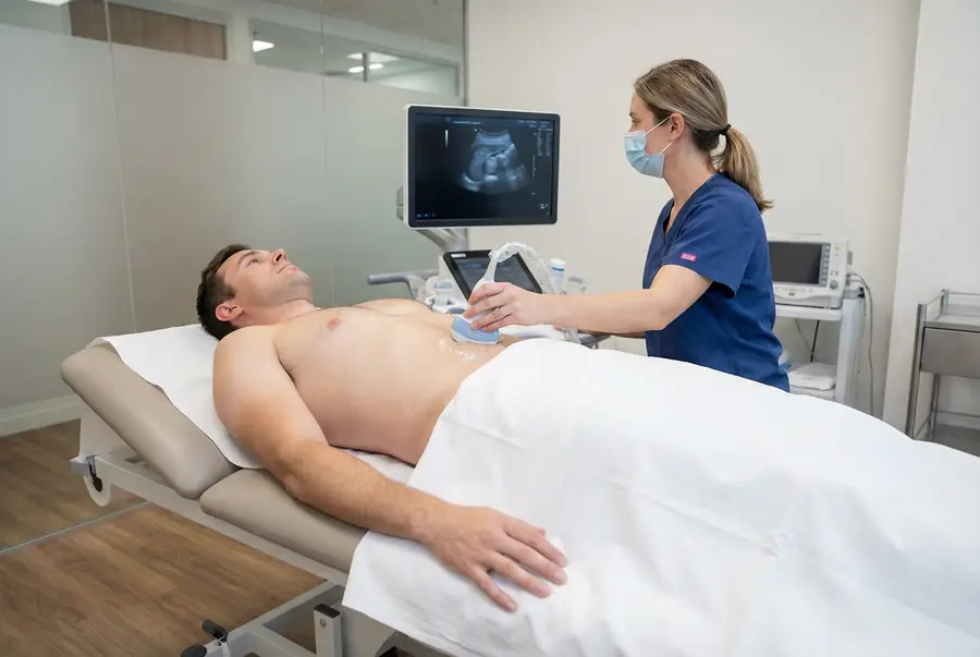
- Gel and scanning: gel is applied and the probe is moved over the skin to obtain vessel images and flow measurements.
- Protocol-specific assessment: gentle compression, a brief breath hold, a change in position or another manoeuvre may be used where appropriate.
- Recording: the examiner saves relevant images, measurements and waveforms.
- Completion: the gel is removed and the team explains the reporting arrangements.
Tell the examiner if probe pressure or positioning causes pain. Any immediate clinical concern is managed through the centre’s escalation process rather than waiting for a routine report.
Benefits, safety, duration and aftercare
Vascular ultrasound provides real-time structural and flow information without ionising radiation. A routine external examination normally requires no injection or sedation and can be repeated when clinically appropriate.
Standard diagnostic ultrasound has no known harmful effects when used appropriately. Probe pressure or compression over a tender area may be uncomfortable, and a reaction to gel is uncommon.
Duration depends on the protocol, the number of vessels and whether one or both limbs are examined. Many appointments take around 30–60 minutes, but your centre will confirm how long to allow. You can usually return to normal activities afterwards unless advised otherwise.
Getting and understanding your results
Your scan images are reviewed by a consultant radiologist or relevant specialist clinician, who prepares a written imaging report. Results are usually available within three working days of the date of your scan. More complex examinations may require additional specialist review and can take slightly longer.
Once your results are available, a post-scan consultation will be arranged with the same medical doctor who conducted your pre-scan consultation. This continuity allows your doctor to consider the findings alongside the symptoms, medical history and concerns you discussed before your scan.
During the consultation, your doctor will explain the report in detail, answer your questions and discuss what the findings mean for you. Where further assessment or care is needed, they will explain the recommended next steps, which may include additional investigations, a specialist referral or follow-up with your usual healthcare team.
How much does a private Doppler ultrasound cost?
Private ultrasound scans with PrivateScans start from £195. This is our general ultrasound starting price, rather than a guaranteed price for every vascular protocol.
The confirmed cost depends on the centre, vessels examined, whether one or both sides are assessed and any specialist requirements. A carotid, arterial, DVT or venous reflux examination may have different arrangements. The service and price will be confirmed before you proceed.
Book a Doppler ultrasound
Your booking includes a 30-minute private telephone consultation with a medical doctor at a convenient time. You can discuss your symptoms, relevant medical history and previous investigations so your doctor can advise on the appropriate examination and urgency.
Where routine outpatient ultrasound is clinically appropriate, a written referral is prepared and the team can help arrange a suitable service, subject to availability. Your report is discussed with the same doctor, who will explain the findings and any necessary next steps. Explore available ultrasound appointments or visit our ultrasound body parts directory.
Possible acute clots, stroke symptoms or sudden limb circulation problems need urgent assessment and should not be booked through a routine pathway.
FAQs
What is the difference between Doppler and duplex?
Doppler assesses blood flow. Duplex combines structural vessel imaging with Doppler information, allowing the examiner to consider the vessel and its flow together.
Are DVT and varicose vein scans the same?
No. A DVT protocol assesses a possible clot, while a reflux examination assesses valve function and maps veins relevant to varicose vein care. The referral should identify the question so the correct protocol is used.
Should I book a routine scan if I think I have a clot?
A possible acute clot needs urgent medical assessment rather than a routine booking. New one-sided leg swelling or pain should be assessed promptly. Chest pain or breathlessness requires emergency assessment.
Does a carotid scan rule out a stroke?
No. It assesses selected neck arteries and does not exclude every cause of neurological symptoms. Suspected stroke symptoms require emergency assessment rather than waiting for a scan.
Do the colours show whether blood flow is healthy?
Not on their own. Colour represents flow according to equipment settings and direction relative to the probe. The clinician interprets the complete examination, including structural images and measurements.
Will I need to fast?
Some abdominal vascular examinations require fasting, while many limb and neck scans do not. Follow the instructions from your centre and ask for advice if fasting could affect a medical condition or medicines.
Can I drive afterwards?
Usually, after a routine external Doppler examination. Follow any individual advice, particularly if your symptoms or another procedure affect your ability to drive safely.
What’s included with your scan?
What is included with your scan:
- A pre-scan telephone consultation with a member of our medical team.
- A written medical referral prepared from the information discussed during your consultation, where clinically appropriate.
- Your scan at the chosen scanning centre.
- A written imaging report prepared by a consultant radiologist or relevant specialist clinician.
- Access to downloadable copies of your scan images where required.
- A post-scan consultation to discuss your results and next steps where needed.