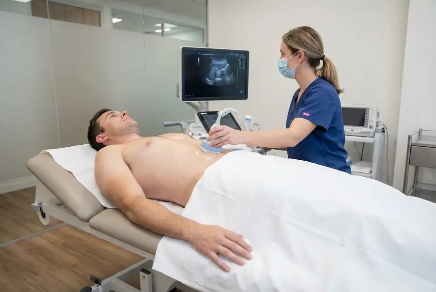
Scan overview
- Abdominal ultrasound uses sound waves to assess selected organs in the abdomen without ionising radiation.
- It may examine the liver, gallbladder, bile ducts, kidneys, spleen and visible parts of the pancreas, according to the referral.
- The scan can identify findings such as gallstones, cysts, some masses, collecting-system dilatation or fluid in the abdomen.
- A routine abdominal examination does not automatically include a full pelvic, bladder, bowel or vascular assessment.
- Preparation depends on the organs being examined. Follow your centre’s instructions about fasting, water and bladder filling.
What is an abdominal ultrasound?
An abdominal ultrasound, also called abdominal sonography, is an external examination of selected organs and structures between the chest and pelvis. A trained clinician moves a handheld probe over the skin to obtain real-time images and relevant measurements.
The technique uses high-frequency sound waves and their returning echoes rather than ionising radiation. It is commonly used as an initial structural investigation when the clinical question is suitable for ultrasound.
The exact coverage depends on your symptoms and referral. An upper abdominal scan, a kidney and bladder scan and a combined abdominal and pelvic examination are different services. Your team will confirm what is included before your appointment.
Why might you need an abdominal ultrasound?
Your doctor may recommend imaging to investigate:
- Persistent or recurrent abdominal discomfort where an organ-related cause is suspected.
- Upper right abdominal pain, including a possible gallbladder concern.
- Abnormal liver blood tests or suspected fatty liver.
- Jaundice or a possible problem with bile drainage.
- A suspected enlarged organ or previously identified structural finding.
- Selected kidney concerns, such as cysts or possible impaired urine drainage.
- Fluid in the abdomen or a finding requiring follow-up.
Not all pain or bloating is explained by ultrasound. Clinical examination, blood tests and other investigations may be needed to identify the cause.
Severe or rapidly worsening abdominal pain, collapse, or pain with fever and feeling acutely unwell needs urgent assessment. New jaundice also requires prompt medical review. Do not wait for a routine private scan when urgent care is needed.
Which organs can be examined?
Liver
The scan can assess liver size, outline and texture, and identify some cysts or focal lesions. Changes may suggest fat accumulation or chronic disease, but blood tests and other assessment are often needed. Read more about liver ultrasound.
Gallbladder and bile ducts
Ultrasound can assess gallstones, gallbladder wall changes and widening of visible bile ducts. It may not show every duct stone or establish the exact cause of obstruction.
Kidneys
The examiner may assess kidney size, cysts, some stones and widening of the collecting system. Ultrasound does not directly measure kidney filtration. A dedicated renal examination may be appropriate for a more specific question.
Spleen and pancreas
Spleen size and selected structural findings can be assessed. The pancreas may be partly obscured by bowel gas, so a normal or limited ultrasound does not exclude pancreatic disease.
Other structures
The abdominal aorta, other vessels or fluid in the abdomen may be assessed where relevant to the referral. Bladder or pelvic imaging needs the appropriate coverage and preparation rather than being assumed to be included.
What findings can abdominal ultrasound identify?
Depending on the area examined, findings may include:
- Gallstones: visible stones within the gallbladder, sometimes with changes requiring clinical assessment.
- Fatty liver: an altered liver appearance consistent with fat accumulation, interpreted alongside risk factors and blood tests.
- Chronic liver changes: selected features that may suggest more advanced disease, although early scarring can be missed.
- Cysts and focal lesions: fluid-filled or solid findings that may be benign or need further imaging.
- Hydronephrosis: widening of the kidney’s collecting system that may suggest impaired drainage.
- Ascites: fluid within the abdominal cavity, which needs assessment of its cause.
- Bile duct dilatation: widening that can prompt further investigation of bile drainage.
Ultrasound may contribute to assessment of inflammation, but it does not establish every infection or diagnose pancreatitis on its own. The clinical picture and laboratory results remain important.
Can ultrasound detect cancer in the abdomen?
Ultrasound can identify some masses or structural abnormalities, but cannot reliably detect or exclude every abdominal cancer. Small lesions, deep structures and areas obscured by bowel gas may be difficult to assess.
Some findings, such as a simple cyst, may have a characteristic benign appearance. Other lesions cannot be confidently classified from ultrasound alone. Fluid-filled does not always mean harmless, and solid does not automatically mean cancer.
Your doctor may recommend additional imaging, such as CT of the abdomen and pelvis or abdominal MRI, specialist review or selected tissue sampling. The choice depends on the finding and the question that still needs answering.
Vascular and specialist ultrasound techniques
Doppler assessment
Doppler techniques add information about blood flow. A dedicated organ vascular or aortic examination has a specific protocol and is not automatically included in a routine abdominal scan.
Elastography
Liver elastography estimates tissue stiffness and may help assess the likelihood of fibrosis. It is different from a routine structural scan and requires confirmation of the appropriate service and interpretation.
Contrast-enhanced ultrasound
In selected specialist settings, an injected ultrasound contrast agent helps characterise a lesion or assess perfusion. This is not routinely required and has separate consent and preparation arrangements.
Ultrasound can also guide biopsy or drainage. These are separate procedures with their own risks, cost and aftercare, rather than automatic components of diagnostic imaging.
What are the limitations of the examination?
Bowel gas, depth, anatomy and the available scanning window can limit visibility. Some organs may be only partly seen, and the report should explain relevant limitations.
A routine abdominal scan is not a comprehensive examination of the stomach or bowel and does not exclude every cause of pain, bleeding or bloating. It also does not directly measure all aspects of organ function: blood and urine tests answer different questions.
A normal scan does not mean continuing or worsening symptoms should be ignored. Your doctor will advise whether further assessment is needed. A dedicated urinary tract or pelvic examination may be more appropriate for some symptoms.
How to prepare for an abdominal ultrasound
Fasting is often requested for an upper abdominal examination, particularly to improve assessment of the gallbladder and relevant surrounding structures. The length of fasting and the advice about water vary between centres and examinations.
Follow the instructions provided for your appointment. Do not assume that all fluids must be avoided. If bladder imaging is included, you may instead need specific drinking and bladder-filling instructions.
Tell the team if you have diabetes, a fluid restriction or medicines that need to be taken with food. Preparation can be adapted where appropriate. Do not stop prescribed medicines without advice.
Wear comfortable clothing that allows access to the abdomen. Bring relevant reports and let the centre know about surgery, pain, mobility needs or a previous reaction to ultrasound gel. Contact the team if preparation instructions are unclear before the day of the scan.
The procedure: what to expect
- Review and consent: the clinician confirms your symptoms, referral and preparation, explains the scan and answers questions.
- Positioning: you usually lie on a couch with the abdomen exposed and appropriate covering. You may be asked to turn onto a side.
- Gel application: water-based gel helps transmit sound between the probe and skin.
- Scanning: the probe is moved across the relevant areas. You may be asked to take a breath and hold it briefly to improve the views.
- Recording: images and measurements are saved, with additional techniques where relevant to the requested examination.
- Completion: the gel is removed and the team explains the reporting arrangements.
You cannot normally hear or feel the sound waves themselves. Probe pressure can be uncomfortable over a tender area; tell the examiner so they can adjust or pause. Any immediate clinical concern is managed through the centre’s escalation process.
Benefits, safety, duration and aftercare
Abdominal ultrasound provides real-time structural information without ionising radiation. A routine examination is external and normally requires no injection or sedation.
Standard diagnostic ultrasound has no known harmful effects when used appropriately. Probe pressure or a full bladder may be uncomfortable, and a skin reaction to the gel is uncommon.
The scan commonly takes around 15–30 minutes, although more extensive or specialist examinations may take longer. You can usually eat, drink, drive and return to normal activities afterwards unless advised otherwise. If you have fasted, you may wish to bring something to eat after the examination.
Getting and understanding your results
Your scan images are reviewed by a consultant radiologist or relevant specialist clinician, who prepares a written imaging report. Results are usually available within three working days of the date of your scan. More complex examinations may require additional specialist review and can take slightly longer.
Once your results are available, a post-scan consultation will be arranged with the same medical doctor who conducted your pre-scan consultation. This continuity allows your doctor to consider the findings alongside the symptoms, medical history and concerns you discussed before your scan.
During the consultation, your doctor will explain the report in detail, answer your questions and discuss what the findings mean for you. Where further assessment or care is needed, they will explain the recommended next steps, which may include additional investigations, a specialist referral or follow-up with your usual healthcare team.
How much does a private abdominal ultrasound cost?
Private ultrasound scans with PrivateScans start from £195. This is our general ultrasound starting price. Your confirmed cost depends on the centre, scan coverage and specialist requirements.
An upper abdominal examination, combined abdominal and pelvic scan, elastography assessment or dedicated vascular study may have different prices. Your consultation helps establish the appropriate investigation, and the examination and cost will be confirmed before you proceed.
Book an abdominal ultrasound
Your booking includes a 30-minute private telephone consultation with a medical doctor at a convenient time. You can discuss symptoms, relevant blood test results, medical history and previous investigations so your doctor can advise whether ultrasound is an appropriate next step.
Where imaging is clinically appropriate, a written referral is prepared and your examination can be arranged at a suitable centre. Your report is discussed with the same doctor, who will explain the findings and any necessary next steps. Explore available ultrasound appointments or return to our ultrasound body parts directory.
FAQs
Can I drink water before the scan?
Follow your centre’s instructions. Fasting may be requested, but advice about water varies. A combined bladder examination may need different preparation. Ask the team if the instructions are unclear rather than assuming that all fluids must be avoided.
Does the scan include every abdominal organ?
Coverage depends on your referral and appointment. Some structures may also be partly obscured. A routine abdominal scan does not automatically include a full pelvic, bowel or vascular examination.
Can ultrasound rule out abdominal cancer?
No. It can identify some suspicious findings but does not detect every cancer. Further imaging or specialist assessment may be needed depending on your symptoms and report.
Can it show gallstones?
Ultrasound can identify many gallbladder stones. It may not show every bile duct stone or establish the cause of all upper abdominal pain. Your doctor will interpret the findings in context.
Does a normal scan mean my liver and kidneys function normally?
Not necessarily. Ultrasound assesses structure. Blood and urine tests are needed to assess many aspects of organ function, and some conditions can be present with a normal-looking scan.
Is elastography included?
Not automatically. Elastography is a specific technique that estimates tissue stiffness, most often for a liver-related question. The team will confirm whether it is appropriate and available.
Can I drive afterwards?
Usually, after a routine diagnostic examination. Follow any individual advice and continue any existing restrictions related to your symptoms or health.
What’s included with your scan?
What is included with your scan:
- A pre-scan telephone consultation with a member of our medical team.
- A written medical referral prepared from the information discussed during your consultation, where clinically appropriate.
- Your scan at the chosen scanning centre.
- A written imaging report prepared by a consultant radiologist or relevant specialist clinician.
- Access to downloadable copies of your scan images where required.
- A post-scan consultation to discuss your results and next steps where needed.