Scan overview
- Ultrasound can assess many accessible lumps close to the skin, providing information about their size, depth and internal appearance.
- It can help distinguish a fluid-filled cyst from a solid lesion, although not every lump can be confidently characterised.
- A general ultrasound is targeted to a specific area or clinical question. It is not a whole-body screening examination.
- Breast, thyroid, testicular and other specialised concerns may need a dedicated examination or specialist pathway.
- The findings help guide care, which may include observation, further imaging or a biopsy. A diagnostic scan does not automatically include treatment.
What is a general body ultrasound?
Ultrasound is an imaging technique that uses high-frequency sound waves to produce real-time pictures of tissues and organs. It has many applications, from examining abdominal organs to investigating a particular lump beneath the skin.
“General ultrasound” describes a range of targeted examinations rather than a single scan that checks the whole body. The appropriate examination depends on where your symptoms are located and what your doctor needs to establish.
For a superficial lump, a trained clinician moves a handheld probe over the area to assess its appearance and relationship to nearby structures. For an internal organ concern, a different probe, technique or preparation may be required. Your consultation helps identify the most suitable examination before booking.
Why might you need an ultrasound of a lump?
Your doctor may recommend ultrasound after examining a lump or reviewing your symptoms. The scan may help answer questions such as:
- Does the palpable lump correspond to a visible soft-tissue lesion?
- How large is it, and how deep beneath the skin does it lie?
- Is it mainly fluid-filled, solid or a mixture of both?
- How does it relate to nearby tissues, a tendon or a gland?
- Are its features sufficiently clear, or is further assessment needed?
Tell your doctor how long the lump has been present, whether it is growing or painful, and whether there has been an injury, infection or previous procedure in the area. A persistent, growing or unexplained lump needs prompt clinical review rather than an assumption that it is harmless.
What can ultrasound show about lumps and bumps?
Ultrasound can describe the location, dimensions, margins and internal characteristics of many accessible soft-tissue findings. Doppler imaging may add information about blood flow when useful.
Some lesions have appearances consistent with a simple cyst, a ganglion or a lipoma. A fluid collection or enlarged lymph node may also account for a palpable swelling. These descriptions help narrow the possibilities, but the diagnosis depends on the full clinical context.
Fluid-filled does not always mean harmless, and solid does not automatically mean cancer. Some findings are uncertain or have features that need specialist assessment. The report should explain the level of confidence and any recommendation for further investigation.
Other uses of general ultrasound
Ultrasound can assess selected internal organs, including the liver, gallbladder, kidneys and pelvic reproductive organs. Depending on the examination, it may identify cysts, some stones, structural changes or findings that need further investigation.
These are distinct examinations with different coverage. A scan of a superficial lump does not automatically include the abdomen or other body areas. You can read about our liver, kidney and pelvic ultrasound examinations for more specific information.
Ultrasound can also guide selected procedures such as biopsy, fluid drainage or an injection. These are separate interventions with their own clinical assessment, consent and aftercare arrangements.
Choosing the right examination
The location of the lump determines the appropriate service. A neck swelling may need a neck ultrasound, a suspected enlarged node may need lymph node assessment, and a concern involving a tendon or muscle may be better addressed by a musculoskeletal examination.
Breast and testicular lumps need an appropriate clinical pathway and should not simply be booked as a generic soft-tissue scan. Other sites or a deep, rapidly growing or otherwise concerning mass may require specialist assessment before imaging.
Your pre-scan consultation provides an opportunity to discuss which scan is likely to answer the clinical question. Where ultrasound is unsuitable, your doctor can explain the recommended next step.
What are the limitations of ultrasound?
Ultrasound cannot reliably confirm or exclude cancer in every lump. Some benign and malignant lesions have overlapping appearances, and a biopsy or specialist review may be needed to establish a diagnosis.
Deep tissue, bone, air and the available scanning window can limit the views. A small or poorly defined lump may not be clearly visible. Ultrasound is also not a comprehensive assessment of bone marrow or every structure within a joint.
If symptoms persist or a lump continues to grow despite a reassuring scan, discuss this with your doctor. Further assessment may include MRI, another imaging examination, sampling or referral to a relevant specialist.
How to prepare for your scan
A superficial soft-tissue ultrasound usually requires little preparation. You can generally eat, drink and take your normal medicines unless the centre gives different instructions.
Wear comfortable clothing that allows access to the area. Bring relevant previous reports and tell the team exactly where you can feel the lump. Let them know about pain, a wound or dressing, mobility needs or a previous reaction to ultrasound gel.
An internal organ examination may require fasting or a full bladder, so follow the instructions for the scan you have booked. If a separate procedure is arranged, its preparation requirements will be explained independently.
The procedure: what to expect
The examiner confirms your symptoms, explains the scan and obtains consent. You are positioned comfortably with the relevant area exposed and your privacy maintained.
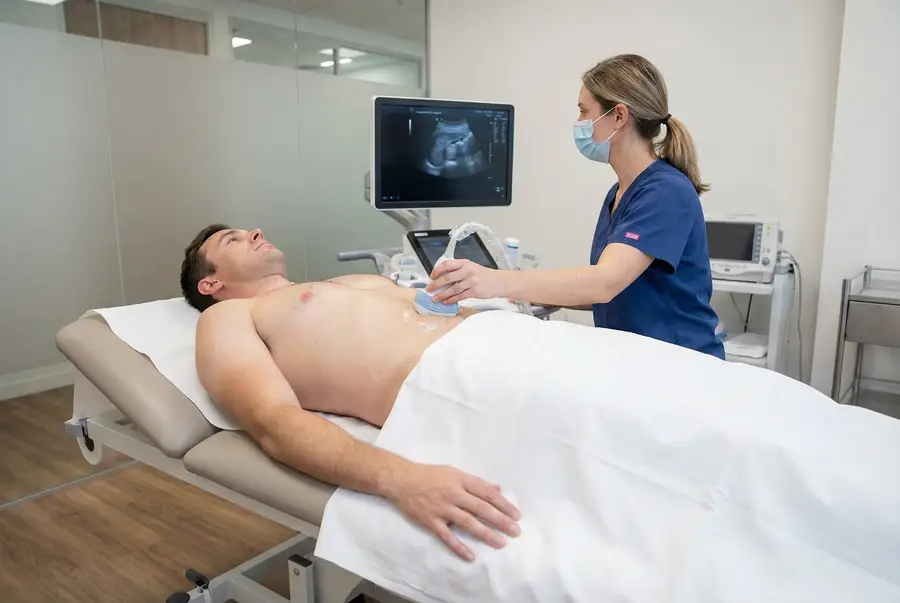
Water-based gel is applied to the skin, and the handheld probe is moved over the lump from different angles. The probe sends sound waves into the tissues and receives returning echoes, which a computer converts into images. Relevant views and measurements are recorded.
You may be asked to point out the lump, change position or move the nearby limb. Probe pressure can be uncomfortable if the area is tender; tell the examiner so they can adjust the technique. Once the scan is complete, the gel is removed and the reporting process is explained.
Safety, duration and aftercare
Standard diagnostic ultrasound uses no ionising radiation and has no known harmful effects when used appropriately. A routine superficial scan normally requires no injection or sedation. A skin reaction to the gel is uncommon.
An appointment often takes around 15–30 minutes, depending on the area and complexity. You can usually drive and return to normal activities afterwards unless advised otherwise.
A diagnostic appointment does not automatically include removal or drainage of a lump. Any treatment or sampling requires a separate decision and consent. Its risks, recovery and reporting arrangements may differ from those of the ultrasound itself.
Getting and understanding your results
Your scan images are reviewed by a consultant radiologist or relevant specialist clinician, who prepares a written imaging report. Results are usually available within three working days of the date of your scan. More complex examinations may require additional specialist review and can take slightly longer.
Once your results are available, a post-scan consultation will be arranged with the same medical doctor who conducted your pre-scan consultation. This continuity allows your doctor to consider the findings alongside the symptoms, medical history and concerns you discussed before your scan.
During the consultation, your doctor will explain the report in detail, answer your questions and discuss what the findings mean for you. Where further assessment or care is needed, they will explain the recommended next steps, which may include additional investigations, a specialist referral or follow-up with your usual healthcare team.
How much does a private ultrasound cost?
Private ultrasound scans with PrivateScans start from £195. This is our general ultrasound starting price. The confirmed price depends on the centre, the area or areas examined and any specialist requirements.
Your medical consultation helps establish the appropriate investigation. Scan coverage and price will be confirmed before you proceed. Biopsy, drainage, injections or other treatments are not automatically included in a diagnostic ultrasound appointment.
Book an ultrasound for a lump or swelling
Your booking includes a 30-minute private telephone consultation with a medical doctor at a convenient time. You can describe the lump, discuss any changes or associated symptoms and review relevant medical history or previous investigations.
Where ultrasound is clinically appropriate, a written referral is prepared and your scan can be arranged at a suitable centre. Your report is discussed with the same doctor, who will explain the findings and any necessary next steps. Explore available ultrasound appointments or visit our ultrasound body parts directory.
FAQs
Is this a whole-body ultrasound?
No. Ultrasound is targeted to the area and clinical question specified in your referral. A scan of a lump does not check all internal organs or screen the whole body for disease.
Can ultrasound tell whether a lump is cancer?
It can identify features that guide the level of concern, but cannot reliably classify every lesion. An uncertain or suspicious finding may require MRI, a biopsy or specialist assessment.
Does a solid lump always mean cancer?
No. Many benign lesions are solid. The examiner considers the overall appearance and your doctor interprets the findings alongside the history and examination.
Will the lump be drained or removed during the scan?
Not as part of a routine diagnostic appointment. Drainage, biopsy or removal requires a separate clinical decision, consent and booking arrangements.
What if the lump does not show on the scan?
Some lumps are difficult to visualise because of their size, depth or similarity to surrounding tissue. A persistent or changing lump still needs clinical review, even if ultrasound is reassuring.
Can I book any lump as a general soft-tissue scan?
The appropriate examination depends on the location and clinical concern. Breast, testicular, thyroid and other specialist concerns may need a dedicated service or referral pathway. Your doctor can advise before booking.
Do I need to fast?
Usually not for a superficial lump examination. Internal organ scans may have different preparation requirements. Follow the instructions supplied for your particular appointment.
What’s included with your scan?
What is included with your scan:
- A pre-scan telephone consultation with a member of our medical team.
- A written medical referral prepared from the information discussed during your consultation, where clinically appropriate.
- Your scan at the chosen scanning centre.
- A written imaging report prepared by a consultant radiologist or relevant specialist clinician.
- Access to downloadable copies of your scan images where required.
- A post-scan consultation to discuss your results and next steps where needed.