Scan overview
- Musculoskeletal (MSK) ultrasound uses sound waves to examine muscles, tendons, ligaments, selected joints and other accessible soft tissues.
- Real-time imaging allows the clinician to assess structures while you move, which can help investigate symptoms such as catching or a tendon moving out of position.
- The examination can assess selected injuries, tendon disorders, fluid collections, inflammation and superficial lumps.
- Ultrasound does not use ionising radiation. A routine diagnostic scan usually needs no injection or sedation and requires little preparation.
- The most appropriate test depends on the area and the clinical question. MRI, X-Ray or CT may be more suitable for structures that ultrasound cannot adequately assess.
What is a musculoskeletal ultrasound scan?
A musculoskeletal ultrasound is a targeted examination of the tissues involved in movement and support. It is commonly used to investigate localised pain, swelling, injury or a palpable lump, and to assess selected changes affecting tendons, muscles, ligaments and joints.
The musculoskeletal system includes bones, muscles, tendons, ligaments, cartilage and other connective tissues. These structures work together to stabilise joints and allow everyday activities such as walking, lifting and reaching. Ultrasound is particularly useful for examining many of the soft tissues close to the skin.
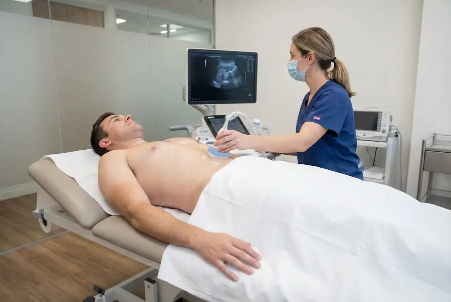
A trained clinician moves a handheld probe over the area of concern. Images appear on a monitor as the examination takes place. Unlike a static image alone, ultrasound can show how some structures behave during movement and allows the examiner to relate an observed change to the location of your symptoms.
Why might you need an MSK ultrasound?
Your doctor may recommend an MSK ultrasound when an examination suggests a problem in a tendon, muscle or other accessible soft tissue. It can support the investigation of:
- Persistent or recurrent pain around a joint.
- Swelling, localised tenderness or a superficial lump.
- A suspected muscle or tendon injury following exercise, sport or another activity.
- Restricted movement, catching, snapping or symptoms that occur in a particular position.
- Possible overuse conditions, such as Achilles tendinopathy, tennis elbow or plantar fasciitis.
- Fluid around a joint or inflammation of a tendon sheath or bursa.
- Selected superficial nerve problems, including changes associated with carpal tunnel syndrome.
- Monitoring a known soft-tissue abnormality when repeat imaging would inform care.
Imaging is considered alongside a clinical assessment. Some symptoms can be managed without a scan, while others require a different examination or urgent assessment. An MSK ultrasound is not a substitute for prompt medical care after a significant injury, for a hot swollen joint with fever, or for sudden loss of movement or sensation.
Which areas can be examined?
MSK ultrasound is a category of targeted examinations rather than a single scan of the whole musculoskeletal system. The area and structures assessed should match your symptoms and the question on your referral.
- Shoulder: selected rotator cuff tendons, the long head of the biceps tendon and surrounding bursae.
- Elbow: accessible tendons, bursae and selected superficial nerves.
- Wrist and hand: tendons, tendon sheaths, ganglion cysts and selected nerve or other soft-tissue abnormalities.
- Hip and groin: selected superficial tendons, bursae and other accessible tissues. A suspected hernia requires an appropriately requested examination.
- Knee: selected superficial tendons and ligaments, joint fluid and cysts, such as a Baker’s cyst behind the knee.
- Ankle and foot: the Achilles tendon, other accessible tendons and ligaments, and the plantar fascia.
- Arms and legs: a particular muscle group, injury or superficial lump.
Ultrasound is not a comprehensive examination of the adult spine, spinal cord or structures deep within a joint. The kidneys, bladder and other internal organs are also outside the scope of a routine MSK appointment.
What can musculoskeletal ultrasound detect?
Tendon and tendon sheath disorders
Ultrasound can assess tendon thickening, altered structure, selected partial or complete tears, and fluid or inflammation around a tendon sheath. These findings may support assessment of conditions such as rotator cuff disease, Achilles tendinopathy or problems affecting the tendons at the elbow.
Muscle injuries
In accessible muscles, ultrasound may show disruption of fibres, a tear or a collection of blood following injury. The examination is tailored to the suspected location and extent of the problem; deep or extensive injuries may need MRI.
Bursitis, joint fluid and inflammation
Bursae are small fluid-containing sacs that reduce friction between tissues. Ultrasound can show an enlarged bursa, a joint effusion or thickened joint lining. Doppler imaging may help assess increased blood flow associated with inflammation. The underlying cause still requires clinical interpretation.
Lumps and selected nerve changes
Ultrasound can help establish whether a superficial lump appears fluid-filled or solid, its size and its relationship to nearby tissues. It can also assess some accessible nerves for enlargement or compression. An uncertain or concerning lump may require further imaging, specialist review or a biopsy; ultrasound cannot classify every mass with certainty.
What are the limitations of MSK ultrasound?
Ultrasound provides detailed images of many superficial structures but does not pass effectively through bone. It cannot comprehensively assess bone marrow, the spinal canal or all of the cartilage and ligaments inside a joint. For example, MRI is often more appropriate for a suspected knee meniscal or cruciate ligament injury.
Image quality depends on the structure’s depth, the available scanning window and the expertise of the examiner. A normal scan does not exclude every injury or explain every cause of pain. Findings such as tendon changes can also be present without symptoms, so they must be interpreted alongside your history and examination.
Ultrasound may demonstrate changes associated with infection or inflammatory disease, but it cannot establish every diagnosis on its own. Blood tests, examination or fluid sampling may be required. It also cannot reliably determine the nature of every tumour or replace appropriate investigation of a growing or otherwise concerning lump.
How does musculoskeletal ultrasound work?
The ultrasound probe, also called a transducer, sends high-frequency sound waves into the tissues and receives the returning echoes. A computer uses these signals to construct images in real time. The sound waves are above the range of human hearing.
A thin layer of water-based gel between the probe and skin helps transmit the sound waves and prevents air from interfering with the images. Different probe types and settings are selected according to the area and the depth of the tissues being examined.
High-frequency probes are commonly used for fine detail near the surface. Doppler settings can assess blood flow when this is relevant, for example around an inflamed tendon or joint lining. The clinician records representative images and measurements for the report.
Assessing tissues during movement
A particular advantage of ultrasound is the ability to observe selected tissues as they move. You may be asked to bend or straighten a joint, rotate a limb or gently contract a muscle while the probe remains over the area.
This dynamic assessment can help demonstrate tendon movement, snapping or impingement that may be less apparent when the joint is still. The clinician may compare the symptomatic area with the opposite side when this would be useful.
Movement is adapted to your comfort and physical ability. You should not force a painful movement, and you can ask the clinician to stop or adjust the examination at any time.
How to prepare for an MSK ultrasound
Most musculoskeletal ultrasound examinations need little preparation. You can usually eat, drink and take your normal medicines as usual. Fasting and a full bladder are not normally necessary for a routine MSK scan, although you should follow any specific instructions from your centre.
Choose comfortable, loose clothing that allows access to the area being scanned. You may need to remove jewellery near the examination site or change into a gown. Bring relevant previous imaging reports and details of any injury, operation or treatment affecting the area.
Tell the team beforehand about mobility difficulties, a wound or dressing, significant tenderness, or any previous reaction to ultrasound gel. If a procedure such as an injection is being considered, separate preparation and consent instructions may apply; this should not be assumed to be part of a diagnostic scan.
The procedure: what to expect
- Clinical review: the examiner confirms your symptoms, the area of concern and the purpose of the scan. They explain the examination and obtain your consent.
- Positioning: you may sit in a chair or lie on a couch, depending on the body part. The area is exposed while your privacy is maintained.
- Gel application: a water-based gel is placed on the skin. It can feel cool when first applied.
- Scanning: the probe is moved across the tissues from different angles. Gentle pressure may be needed to obtain images, and an already tender area can feel uncomfortable.
- Movement and measurements: where appropriate, you are asked to move the joint or limb. Images and measurements are recorded, with Doppler assessment when useful.
- Completion: the gel is wiped away and the team explains the reporting arrangements.
Tell the examiner if pressure or positioning causes pain. They can adjust the technique or pause the examination. Any immediate clinical concern is managed through the centre’s escalation process; your formal findings are explained through your report and follow-up consultation.
Benefits, safety and possible discomfort
MSK ultrasound offers real-time assessment without ionising radiation. It can focus closely on a symptomatic area, assess selected tissues during movement and provide useful information about fluid, inflammation and superficial soft-tissue changes.
Routine diagnostic ultrasound has no known harmful effects when used appropriately. It normally requires no contrast injection or sedation. Pressure over an injury may cause temporary discomfort, and a skin reaction to the gel is uncommon. Let the team know if you have a known sensitivity.
Ultrasound can also guide certain injections, aspirations or biopsies. These are separate procedures with their own benefits, risks and consent requirements. Their availability and cost should be confirmed independently rather than assumed to be included with your diagnostic appointment.
When might another scan be more appropriate?
Your clinician will recommend imaging based on the structures that need assessment and the information required to guide care. Depending on your symptoms, this may include:
- MRI: detailed assessment of deeper soft tissues, bone marrow and many structures inside joints.
- X-Ray: assessment of fractures, alignment and selected changes affecting bones and joints.
- CT: detailed bone imaging, particularly for complex fractures or anatomy that needs closer assessment.
Occasionally, a specialist may advise another investigation, such as a nuclear medicine bone scan, where the clinical question warrants it. A combination of tests may be needed. The purpose of your pre-scan consultation is to help select an appropriate investigation rather than proceeding with a scan that is unlikely to answer the question.
How long does the scan take, and what happens afterwards?
A targeted MSK ultrasound commonly takes around 20–30 minutes. The duration varies with the area, the number of structures assessed and the complexity of the findings. Allow additional time for registration, changing and any other appointment requirements.
After a routine diagnostic scan, you can usually drive, return to work and continue normal activities. The examination itself does not normally require recovery time. Follow any existing advice about your injury or activity restrictions; having a scan does not mean that it is safe to resume sport or increase loading.
Getting and understanding your results
Your scan images are reviewed by a consultant radiologist or relevant specialist clinician, who prepares a written imaging report. Results are usually available within three working days of the date of your scan. More complex examinations may require additional specialist review and can take slightly longer.
Once your results are available, a post-scan consultation will be arranged with the same medical doctor who conducted your pre-scan consultation. This continuity allows your doctor to consider the findings alongside the symptoms, medical history and concerns you discussed before your scan.
During the consultation, your doctor will explain the report in detail, answer your questions and discuss what the findings mean for you. Where further assessment or care is needed, they will explain the recommended next steps, which may include additional investigations, a specialist referral or follow-up with your usual healthcare team.
How much does a private musculoskeletal ultrasound cost?
Private ultrasound scans with PrivateScans start from £195. This is our general ultrasound starting price. Your confirmed cost depends on the scanning centre, the body area, the scope of the examination and any specialist requirements.
A scan of one joint or tendon is different from assessment of several areas. Ultrasound-guided treatment or sampling is not automatically included in a diagnostic scan. Your medical consultation helps clarify the appropriate examination, and its coverage and price will be confirmed before you proceed.
Book a musculoskeletal ultrasound
Your booking includes a 30-minute private telephone consultation with a medical doctor, arranged at a convenient time. This gives you the opportunity to describe your symptoms, discuss previous injuries or investigations and ask whether ultrasound is an appropriate next step.
Where clinically appropriate, a medical referral is prepared and your examination can be arranged at a suitable centre. After the scan, your report is discussed with the same doctor, who can explain the findings in context and advise on any necessary next steps. Explore available ultrasound appointments or return to our ultrasound body parts directory.
FAQs
What is the musculoskeletal system?
It is the system of bones, muscles, tendons, ligaments, joints, cartilage and connective tissues that supports your body and enables movement. An MSK ultrasound focuses on selected tissues in a particular area rather than examining the entire system.
Do I need an existing referral?
You can contact PrivateScans without obtaining a GP referral beforehand. Your included medical consultation assesses whether the proposed examination is appropriate, and a written referral is prepared where clinically appropriate. Any centre-specific requirements are confirmed before your appointment.
Who performs the scan?
Your examination is carried out by an appropriately trained imaging professional, such as a sonographer or radiologist with relevant MSK expertise. Other suitably trained clinicians may perform specialist ultrasound examinations in some settings. A written report is prepared by the relevant reporting clinician.
Can ultrasound show a torn tendon or muscle?
It can identify many tears in accessible tendons and muscles, and may help assess their extent. The ability to detect an injury depends on its location and the tissues involved. MRI may be advised when a structure is too deep, the findings are uncertain or a wider assessment is needed.
Can MSK ultrasound assess the spine?
A routine MSK ultrasound does not comprehensively assess the adult spine, spinal canal, discs or spinal cord. Depending on the symptoms, MRI, X-Ray or CT may be more suitable. Selected superficial tissues near the spine can sometimes be examined for a specific clinical question.
Does my scan include an injection?
No injection is normally needed for a diagnostic MSK ultrasound. Ultrasound-guided injections or other procedures require a separate clinical decision, consent and booking arrangements. The team will confirm what is included in your appointment.
Can I exercise after the examination?
There is usually no restriction caused by the diagnostic scan itself. However, continue to follow the advice you have been given about your symptoms or injury. Your doctor can discuss the findings and whether further assessment is needed before you return to a particular activity.
What’s included with your scan?
What is included with your scan:
- A pre-scan telephone consultation with a member of our medical team.
- A written medical referral prepared from the information discussed during your consultation, where clinically appropriate.
- Your scan at the chosen scanning centre.
- A written imaging report prepared by a consultant radiologist or relevant specialist clinician.
- Access to downloadable copies of your scan images where required.
- A post-scan consultation to discuss your results and next steps where needed.