Scan overview
- A bladder ultrasound uses sound waves to produce real-time images of the bladder, without ionising radiation.
- The scan is performed externally over the lower abdomen, usually with a full bladder. You may also be asked to empty your bladder for a second set of measurements.
- It can assess bladder size, wall appearance, stones and some abnormal growths, and measure how much urine remains after urination.
- Ultrasound findings are interpreted alongside your symptoms, medical history and other tests. A normal scan does not exclude every cause of urinary symptoms.
- Your booking includes medical consultation support before your scan and a follow-up discussion of your report.
What is a bladder ultrasound?
A bladder ultrasound is a non-invasive examination that helps clinicians assess the structure of the bladder and how effectively it empties. It is sometimes called a bladder sonogram or a transabdominal bladder scan.
The bladder is a muscular organ in the pelvis that stores urine produced by the kidneys. Urine travels from each kidney through a tube called a ureter, collects in the bladder and leaves the body through the urethra. Problems affecting any part of this system can cause similar symptoms, so the area scanned is guided by the clinical question.
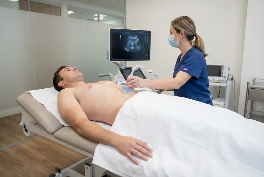
A sonographer or another appropriately trained clinician moves a handheld ultrasound probe across the skin of your lower abdomen. Images appear on a screen as the examination takes place. Measurements taken before and, where indicated, after urination can help assess bladder filling and emptying.
Why might you need a bladder ultrasound?
Your clinician may recommend a bladder ultrasound to investigate persistent or recurrent urinary symptoms, assess a known bladder problem or check for incomplete emptying. Reasons for assessment can include:
- Difficulty starting urination, a weak stream or a feeling that the bladder has not emptied fully.
- Urinary frequency, urgency, leakage or difficulty controlling urination.
- Recurrent urinary tract infections.
- Pain or discomfort when passing urine.
- Blood in the urine, as part of a wider clinical investigation.
- Monitoring a previously identified structural abnormality.
The scan is selected following clinical assessment; not every urinary symptom requires imaging. Urine tests, examination and other investigations may be more appropriate or may be needed alongside ultrasound.
If you suddenly cannot pass urine, seek urgent medical assessment rather than waiting for a routine scan. Blood in the urine also requires prompt medical review, even if it happens only once.
What can a bladder ultrasound show?
A bladder ultrasound can identify structural changes and provide useful information about bladder emptying. Findings that may be assessed include:
- Urinary retention: measuring urine remaining after you have passed urine helps identify incomplete emptying. Further assessment is needed to establish the cause.
- Bladder stones: mineral deposits within the bladder can sometimes contribute to irritation, infection or difficulty passing urine.
- Bladder wall changes: thickening or an irregular appearance may be associated with inflammation, longstanding obstruction or other conditions. The degree of bladder filling affects how the wall appears.
- Bladder diverticula: pouches extending from the bladder wall may be visible and can sometimes be associated with urinary stasis or recurrent infection.
- Abnormal growths: some masses projecting into the bladder can be seen, but further investigation is usually necessary to determine their nature.
- Selected anatomical abnormalities: for example, a ureterocele, where the end of a ureter forms a bulge within the bladder.
These findings are considered with your symptoms and other results. Ultrasound alone cannot reliably distinguish every benign change from cancer, and it does not replace cystoscopy or tissue sampling when these are clinically indicated.
What are the limitations of the scan?
Ultrasound does not detect every bladder abnormality. Small lesions and flat changes in the bladder lining may not be visible, and image quality can be affected by insufficient bladder filling, bowel gas or individual anatomy. A reassuring scan therefore does not rule out bladder cancer or remove the need to investigate continuing symptoms.
A urinary tract infection is generally assessed using symptoms and urine testing; ultrasound may look for an underlying structural problem in selected cases. Likewise, the scan can show incomplete emptying associated with neurological conditions, but cannot by itself diagnose a neurogenic bladder or fully assess bladder pressure and function.
Routine bladder ultrasound does not reliably establish whether urine refluxes towards the kidneys. Specialist investigations may be needed for suspected reflux. Your clinician may also recommend kidney or urinary tract imaging, urine tests, cystoscopy, CT, MRI or urodynamic testing, depending on the findings and clinical concern.
Types of bladder ultrasound
Transabdominal ultrasound
This is the usual outpatient examination. The probe is placed over the lower abdomen to assess the filled bladder, its wall and visible contents. There is no internal instrument involved in a standard transabdominal scan.
Post-void residual measurement
After the initial images, you may be asked to use the toilet and then return for another measurement. This estimates the volume of urine left in the bladder. The result is interpreted in context, including how full your bladder was and whether you were able to urinate normally during the appointment.
Doppler assessment
Colour Doppler may be used when helpful to assess blood flow in a visible structure or observe urine entering the bladder from the ureters. It supplements the standard images and is not a complete test of urinary tract function.
Other specialist applications
Portable bedside scanners can estimate bladder volume in acute care. Three-dimensional imaging, contrast-enhanced techniques and ultrasound used during surgery have more specialised applications. They are not routine components of a standard private bladder ultrasound and would require separate clinical consideration.
Bladder ultrasound or a wider urinary tract scan?
A bladder examination focuses on the bladder and, where requested, residual urine after emptying. A wider urinary tract ultrasound may also assess the kidneys. A pelvic or prostate examination has a different clinical focus and should not be assumed to be included in every bladder appointment.
During your pre-scan consultation, your doctor will consider the symptoms you describe and advise which examination is appropriate. The scan coverage and any additional requirements will be confirmed before your appointment. You can also explore our ultrasound body parts directory for information about other examinations.
How does bladder ultrasound work?
The ultrasound probe, also called a transducer, sends high-frequency sound waves into the body and receives the echoes reflected from tissues and fluid boundaries. A computer converts these signals into images that can be viewed in real time.
A water-based gel helps sound waves pass between the probe and your skin. You cannot usually hear or feel the sound waves themselves. Unlike CT and X-Ray examinations, ultrasound does not use ionising radiation.
The equipment typically includes a monitor, a control console and a handheld probe. The clinician adjusts the settings, captures representative images and takes measurements relevant to the reason for your scan.
How to prepare for a bladder ultrasound
A full bladder usually provides clearer images and allows useful measurements. Your scanning centre will give you specific drinking instructions and tell you when to avoid passing urine before the examination. Instructions may include drinking approximately 500 ml of water before arrival, but the amount and timing can vary.
Follow the instructions supplied for your appointment rather than relying on a general guide. If you have a fluid restriction, heart or kidney problems affecting fluid intake, a catheter or difficulty holding urine, contact the team beforehand so preparation can be adapted. Tell the team if filling your bladder becomes painful; do not continue drinking beyond what you have been advised.
Wear comfortable clothing that allows access to the lower abdomen. Take your usual medicines unless advised otherwise. Fasting is not normally needed for a bladder-only scan, although a combined examination may have different requirements. Bring any relevant previous reports and let the team know about mobility or communication needs.
The procedure: what to expect
- Review and consent: your clinician confirms the reason for the examination, checks preparation and explains what will happen. You can ask questions before proceeding.
- Positioning: you usually lie on your back on an examination couch. Your lower abdomen is exposed while the rest of your body remains covered as appropriate.
- Gel and scanning: gel is applied to the skin and the probe is moved over the lower abdomen with gentle pressure. A full bladder can make this feel uncomfortable; tell the clinician if you need a pause.
- Images and measurements: the clinician records the bladder from different angles and measures relevant structures. You may be asked to change position or briefly hold your breath.
- Emptying assessment: if a post-void measurement is required, you use the toilet and return promptly for a further scan.
- Completion: the gel is wiped away and you can dress. The team explains the reporting process and any immediate instructions.
An examination may be modified to suit your symptoms or physical needs. Any urgent finding is managed through the centre’s clinical escalation process; routine findings are explained through your formal report and follow-up consultation.
Benefits, safety and possible discomfort
Bladder ultrasound provides real-time information without an injection, an incision or ionising radiation. It is an established method for assessing bladder appearance and measuring residual urine, and you can usually return to normal activities immediately afterwards.
Standard diagnostic ultrasound has no known harmful effects when used appropriately. The main source of discomfort is usually the full bladder or pressure from the probe, particularly if you already have pelvic pain. Let the clinician know so that they can adjust the examination.
No sedation or contrast agent is normally required for a routine transabdominal bladder scan. If you are pregnant or think you may be, tell the team so they have a complete clinical history and can plan your care appropriately.
How long does it take, and what happens afterwards?
The scan itself commonly takes around 10–20 minutes. Allow extra time for registration, preparation and any additional bladder filling or post-void assessment. Your appointment confirmation will indicate how long to allow at your chosen centre.
You can normally eat, drink, drive and return to work or your usual activities after a standard bladder ultrasound. You can empty your bladder once the clinician confirms that the necessary images have been obtained. Further preparation or aftercare instructions may apply if another examination is being performed at the same appointment.
Getting and understanding your results
Your scan images are reviewed by a consultant radiologist or relevant specialist clinician, who prepares a written imaging report. Results are usually available within three working days of the date of your scan. More complex examinations may require additional specialist review and can take slightly longer.
Once your results are available, a post-scan consultation will be arranged with the same medical doctor who conducted your pre-scan consultation. This continuity allows your doctor to consider the findings alongside the symptoms, medical history and concerns you discussed before your scan.
During the consultation, your doctor will explain the report in detail, answer your questions and discuss what the findings mean for you. Where further assessment or care is needed, they will explain the recommended next steps, which may include additional investigations, a specialist referral or follow-up with your usual healthcare team.
How much does a private bladder ultrasound cost?
Private ultrasound scans with PrivateScans start from £195. This is our general ultrasound starting price; the confirmed cost depends on the scanning centre, the examination required and whether additional areas or specialist assessments are included.
Your medical consultation helps establish the appropriate scan coverage before you proceed. You will receive confirmation of the examination and its price before booking. A combined kidney and bladder examination, for example, may have a different price from a bladder-only scan.
Book a bladder ultrasound
If you have ongoing urinary symptoms, a clinical assessment can help establish whether a bladder ultrasound is the right next investigation. Your included pre-scan telephone consultation gives you the opportunity to discuss your concerns, relevant medical history and previous investigations with a medical doctor.
Where imaging is appropriate, we can help arrange your scan at a suitable centre, followed by a discussion of the report and any recommended next steps. Start by exploring available ultrasound appointments. Routine private scanning is not a substitute for urgent assessment if you suddenly cannot pass urine or become acutely unwell.
FAQs
Do I need a full bladder?
Usually, yes. A filled bladder is easier to assess and helps the clinician obtain useful images and measurements. Follow your centre’s drinking instructions and contact the team if you have a fluid restriction, a catheter or difficulty holding urine.
Will I need to empty my bladder during the appointment?
You may be asked to urinate after the first images, then return for a post-void residual measurement. This shows how much urine remains and helps assess emptying. It is performed when relevant to your symptoms or referral.
Can a bladder ultrasound diagnose cancer?
Ultrasound can identify some bladder masses, but it cannot reliably confirm or exclude cancer. Small or flat abnormalities may be missed. Cystoscopy and, where appropriate, biopsy may be needed, particularly when blood in the urine or other concerning symptoms require investigation.
Can ultrasound diagnose a urinary tract infection?
A UTI is usually assessed from your symptoms and urine testing. Ultrasound may be requested to look for stones, retention or structural abnormalities in selected people, such as those with recurrent infections. It does not replace urine tests.
Is the scan internal or painful?
A standard bladder ultrasound is performed externally over the lower abdomen. The probe pressure and a full bladder can cause discomfort, but no internal instrument is used. Tell the clinician if you are in pain so they can adapt the examination.
Are my kidneys included in a bladder scan?
Not automatically. A urinary tract examination may include both kidneys and the bladder, while a bladder-only scan has a narrower focus. Your doctor will advise on appropriate coverage, and the examination will be confirmed before your appointment.
What if my scan is normal but symptoms continue?
Discuss ongoing symptoms with your doctor. A normal ultrasound does not exclude every urinary condition. Further urine tests, cystoscopy, other imaging or functional bladder testing may be advised depending on your symptoms and clinical history.
What’s included with your scan?
What is included with your scan:
- A pre-scan telephone consultation with a member of our medical team.
- A written medical referral prepared from the information discussed during your consultation, where clinically appropriate.
- Your scan at the chosen scanning centre.
- A written imaging report prepared by a consultant radiologist or relevant specialist clinician.
- Access to downloadable copies of your scan images where required.
- A post-scan consultation to discuss your results and next steps where needed.