Scan overview
- A kidney CT, also called a renal CT, produces detailed cross-sectional images of the kidneys and relevant urinary tract structures.
- It may help assess stones, a cyst or mass, a drainage concern or selected complications.
- A non-contrast stone scan, renal mass examination, CT urography and renal CT angiography use different protocols.
- CT uses X-rays. Contrast suitability is assessed separately, including kidney function where relevant.
- PrivateScans CT scans start from £395, with the final price confirmed for your chosen centre and examination.
What is a kidney CT scan?
A kidney CT is a computed tomography examination planned to investigate a specific renal or urinary tract concern. It combines X-ray measurements taken from different angles to create detailed cross-sectional images.
The kidneys sit towards the back of the abdomen and filter waste from the blood while helping regulate fluid, electrolytes and blood pressure. Urine drains from each kidney through a ureter to the bladder.
CT can assess kidney anatomy and selected abnormalities. The area covered and use of contrast depend on the question: a scan for a suspected stone is different from an examination designed to characterise a mass or assess the urinary tract lining.
The findings are interpreted alongside symptoms, clinical examination, blood and urine tests and previous imaging. CT does not directly measure every aspect of kidney function or replace those other assessments.
Why might a kidney CT be recommended?
Your clinician may recommend CT where a structural concern needs investigation or a previous test has left an unanswered question. The protocol and urgency depend on the suspected condition.
Reasons may include:
- Suspected urinary stones or a defined concern following a previous stone episode.
- Blood in the urine requiring an appropriate urinary tract assessment.
- A cyst or mass needing further characterisation after ultrasound or another test.
- Possible obstruction or a dilated collecting system.
- Selected complications of infection, injury or previous treatment.
- A specific question about renal blood vessels.
Flank pain, recurrent urinary symptoms or abnormal kidney blood tests can have several causes. They do not automatically mean CT is the best first investigation.
Severe pain with fever, inability to pass urine or marked illness needs prompt medical assessment. A suspected infected obstruction is an urgent concern; do not wait for a routine private scan.
What can a kidney CT help assess?
Kidney and ureteric stones
A dedicated non-contrast examination can identify many urinary stones and assess their size and location, together with selected signs of obstruction. The interpretation considers the symptoms and anatomy because other calcifications may lie near the urinary tract. No test is perfect, and persistent symptoms may need further review even after imaging.
Cysts and complex cystic abnormalities
Many simple kidney cysts are benign and adequately assessed with ultrasound or other imaging. CT with an appropriate protocol may help characterise a complex cyst, including its walls, internal components and enhancement where contrast is used.
Renal masses
CT can help assess the location, size and appearance of a mass and its relationship to surrounding tissues. A dedicated contrast protocol may be needed. Imaging cannot definitively classify every mass as benign or malignant, and specialist review or further tests may be required.
Hydronephrosis and possible obstruction
Hydronephrosis describes dilatation of the urine-collecting system. CT may help identify a possible cause or level of obstruction, depending on the protocol. Dilatation alone does not establish every aspect of drainage function.
Infection and abscesses
Where clinically indicated, CT may assess a suspected complication such as an abscess or another concern in someone with an infection. Most urinary infections do not require CT. Clinical assessment, urine tests and treatment should not be delayed while arranging routine imaging.
Multiple cysts and known kidney conditions
Selected examinations may assess anatomy or a particular complication in someone with an established condition, including polycystic kidney disease. CT does not predict kidney failure from appearance alone or replace ongoing kidney function monitoring.
Trauma and postoperative findings
CT may help assess selected injuries or a specific concern after treatment. Acute trauma needs appropriate urgent assessment, with the protocol selected in that setting. Previous operation details and earlier imaging assist interpretation of postoperative changes.
Blood vessels and other urinary structures
Dedicated vascular imaging or CT urography may address particular questions about renal vessels, ureters or the collecting systems. A routine renal scan does not automatically include a complete assessment of every urinary structure or blood vessel.
Different kidney CT protocols
Non-contrast CT for stones
A stone-focused examination, sometimes described as a CT KUB, assesses the kidneys, ureters and bladder region according to the protocol. It usually does not need an intravenous contrast injection.
Contrast-enhanced renal CT
An iodine-based injection may be used when assessing a mass or another renal concern. Images are timed to show how tissues enhance. Some questions need acquisitions at more than one phase, but additional phases are selected only where useful.
CT urography
CT urography uses a tailored protocol to assess the urinary tract. It may include delayed images after contrast to show the collecting systems and ureters. It does not replace every investigation of the bladder lining, and other assessment may still be required.
Renal CT angiography
This is a dedicated vascular examination for a specific concern about the renal blood vessels. It is not automatically included in a standard stone or mass investigation.
Multiphase imaging and reconstructions
Terms such as triphasic CT refer to particular acquisition protocols. The number and timing of phases depend on the referral. Additional views and selected 3D reconstructions may support planning, but do not mean that every test is included in one booking.
Does the scan include the bladder and ureters?
The coverage depends on the referral. A stone protocol or CT urography may include the urinary tract through to the bladder, while a focused renal mass examination may have a different emphasis.
Even where a structure is visible, the scan may not provide every specialised assessment of it. For example, CT does not replace cystoscopy or every other investigation recommended for blood in the urine.
Tell your clinician about the full pattern of symptoms and previous findings. The team will clarify the required protocol before booking rather than assume that every kidney CT is interchangeable.
Kidney CT, ultrasound and MRI
Ultrasound is useful for many initial kidney and urinary tract questions and does not use ionising radiation. CT can add detailed information about stones, a mass or selected complications where indicated.
A kidney MRI may help characterise a particular lesion or address another specialist concern without ionising radiation. It is not automatically a substitute for every CT protocol, particularly a stone investigation.
Blood and urine tests remain important for kidney function and infection assessment. The most useful combination of tests depends on the clinical question, your suitability and previous results.
Contrast and kidney function
Some examinations use an iodine-based contrast injection through a cannula in a vein. Many stone-focused scans do not require it. The need for contrast should be determined by the question being investigated.
Tell the team about kidney disease, a recent deterioration in kidney function, dialysis, previous contrast reactions and all regular medicines. A recent kidney function blood test may be required before the protocol is confirmed.
Reduced kidney function and a previous contrast reaction are separate considerations. The team assesses each individually and may adapt the examination or recommend another approach.
An injection can cause a brief warm feeling or metallic taste. Reactions can occur; tell the team immediately if you feel unwell, develop breathing difficulty or experience other concerning symptoms.
Follow the centre’s specific instructions about medicines, including metformin where relevant. Do not stop treatment or increase fluid intake beyond existing restrictions without clinical advice.
How does renal CT work?
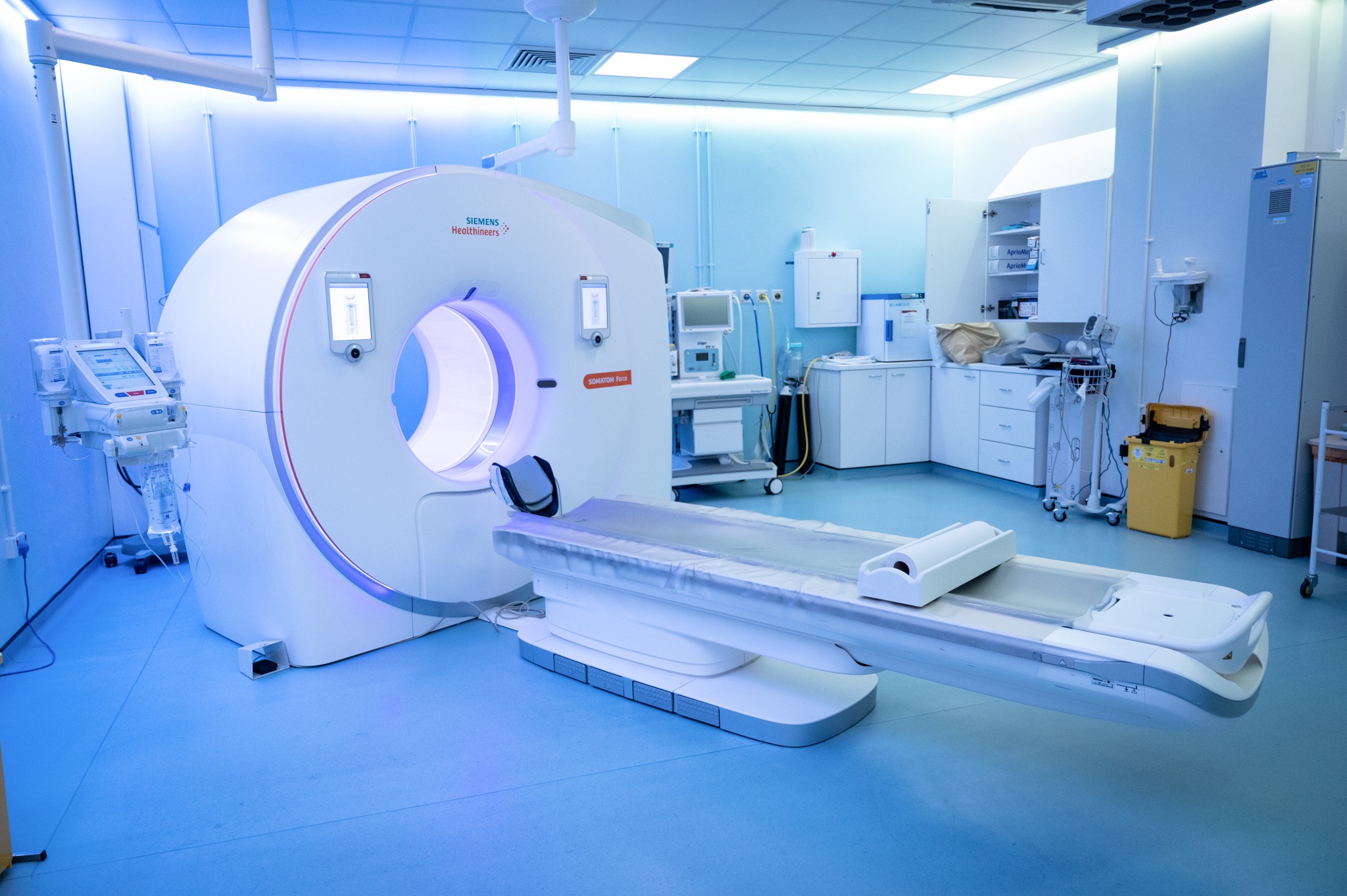
A CT scanner has a short, ring-shaped opening and a motorised table. An X-ray source and detectors rotate around the area being examined, and a computer processes the measurements into cross-sectional images.
The radiologist can review the images in several planes using display settings appropriate to the tissue and clinical question. If contrast is used, images may be acquired at specific times after the injection.
CT does not use a strong magnetic field for imaging. Image acquisition is usually brief, but remaining still and following any short breathing instructions helps reduce blurring. The radiographer can see and communicate with you throughout.
Preparing for your kidney CT
Your centre will provide instructions for the required protocol. Food, fluid and bladder preparation can vary. Follow those instructions rather than assuming that every kidney CT needs the same preparation.
Before your appointment, disclose:
- Pregnancy or possible pregnancy.
- Kidney disease, dialysis, transplantation or a recent change in kidney function.
- A previous reaction to CT contrast.
- Relevant medical conditions and all regular medicines.
- Previous urinary tract surgery and relevant earlier imaging.
- Difficulty lying flat or holding your breath briefly.
Bring requested blood test results and relevant reports. Ask for advice if fasting could affect diabetes treatment or another condition. Do not alter medicines or prescribed fluid restrictions without clinical guidance.
You may change into a gown and remove metal items near the scanned area to avoid image distortion. CT does not have the same magnetic restrictions as MRI.
What happens during the examination?
- Review and explanation. The team checks your referral, preparation, relevant blood results and any contrast requirements.
- Preparing for imaging. You may change clothing and remove items near the area that could affect image quality.
- Positioning. You usually lie on your back on the scanner table. Tell the radiographer if a position is uncomfortable.
- Image acquisition. The table moves through the short scanner opening while images are obtained. Keep still and follow any breathing instructions.
- Contrast and additional phases if required. An intravenous injection is given and further images are timed according to the protocol. Delayed images may extend the examination.
- Completion. The radiographer checks image quality and explains any observation or aftercare instructions.
Tell the team if you feel unwell or need help. The radiographer obtains the images; a radiologist subsequently interprets them and prepares the report.
Benefits, radiation and limitations
Renal CT can provide detailed anatomical information quickly and support decisions about a defined urinary tract concern. A routine examination does not involve an incision.
CT uses ionising radiation. The expected benefit and exposure are considered before scanning, and the protocol is selected to obtain useful information while managing exposure. Multiple phases are not necessary for every question. Tell the team about pregnancy or possible pregnancy beforehand.
Contrast has additional suitability considerations and potential side effects, including kidney-related considerations in relevant patients. These are assessed before an injection is recommended.
CT does not explain every urinary symptom or directly replace kidney function testing. Uncertain findings may need specialist review or further investigation rather than a definitive diagnosis from the images alone.
Appointment length and aftercare
A routine appointment often takes around 15 to 30 minutes, but preparation, contrast, delayed phases or observation can add time. Your centre will confirm the expected length for your protocol.
Most people can return to their usual routine after a standard examination. Continue any existing advice about your condition and medicines unless the clinical team gives different instructions.
If contrast is used, follow the centre’s observation and aftercare advice. Fluid instructions should take account of kidney or heart conditions and any prescribed restrictions. Do not assume that everyone should drink extra water.
Getting and understanding your results
Your scan images are reviewed by a consultant radiologist or relevant specialist clinician, who prepares a written imaging report. Results are usually available within three working days of the date of your scan. More complex examinations may require additional specialist review and can take slightly longer.
Once your results are available, a post-scan consultation will be arranged with the same medical doctor who conducted your pre-scan consultation. This continuity allows your doctor to consider the findings alongside the symptoms, medical history and concerns you discussed before your scan.
During the consultation, your doctor will explain the report in detail, answer your questions and discuss what the findings mean for you. Where further assessment or care is needed, they will explain the recommended next steps, which may include additional investigations, a specialist referral or follow-up with your usual healthcare team.
How much does a private kidney CT cost?
PrivateScans CT scans start from £395. This is our general CT starting price. Your final cost depends on the chosen centre, required coverage and whether contrast or a specialist protocol is needed.
A stone scan, renal mass examination, CT urography and vascular study may involve different arrangements and pricing. Our team will confirm the examination, price and included services before you proceed.
Arrange a private kidney CT
Your booking includes a 30-minute private telephone consultation with a medical doctor at a convenient time. The doctor will discuss your symptoms, kidney health, previous investigations, medicines and treatment before advising whether renal CT is clinically appropriate.
Where imaging is indicated, the referral identifies the clinical question and relevant findings. This helps the scanning team plan the protocol and gives the radiologist useful context.
You can explore our other CT body part examinations. Acute concerning symptoms need prompt clinical care rather than waiting for a routine scan.
FAQs
Will I need contrast for a kidney stone scan?
A dedicated stone protocol usually does not require intravenous contrast. Other renal questions may need it, so the referral determines the protocol.
Can CT miss a kidney stone?
CT detects many urinary stones, but no test is perfect. Stone size, composition, protocol and image quality can affect assessment. Persistent or worsening symptoms need clinical review rather than relying on a scan alone.
Does every kidney CT include the bladder?
No. Coverage and sequences depend on the question. CT urography and a stone-focused examination differ from a dedicated renal mass protocol.
Can I have contrast if I have kidney disease?
This needs individual assessment. Tell the centre about your kidney health and provide blood results if requested. The team will decide whether contrast is appropriate or another approach is needed.
What is a multiphase kidney CT?
It obtains images at selected times around a contrast injection to answer a particular question. Not every renal examination needs multiple phases, and the protocol is tailored to the referral.
Can CT measure kidney function?
CT primarily assesses structure and selected enhancement or drainage patterns. Blood and urine tests remain important for measuring and monitoring kidney function.
When will my results be available?
Results are usually available within three working days of your scan. More complex examinations can take slightly longer. A post-scan consultation will be arranged with the same doctor who conducted your pre-scan consultation to explain the findings and any necessary next steps.
What’s included with your scan?
What is included with your scan:
- A pre-scan telephone consultation with a member of our medical team.
- A written medical referral prepared from the information discussed during your consultation, where clinically appropriate.
- Your scan at the chosen scanning centre.
- A written imaging report prepared by a consultant radiologist or relevant specialist clinician.
- Access to downloadable copies of your scan images where required.
- A post-scan consultation to discuss your results and next steps where needed.