Scan overview
- An ankle CT provides detailed images of the bones and joint surfaces around the ankle.
- It may help assess complex fractures, alignment, arthritis, osteochondral abnormalities and selected postoperative concerns.
- MRI or ultrasound may be more appropriate where a ligament, tendon or other soft tissue injury is the main concern.
- CT uses X-rays. The expected clinical benefit and radiation exposure are considered before scanning.
- PrivateScans CT scans start from £395, with the final price confirmed for your chosen centre and examination.
What is an ankle CT scan?
An ankle CT is a computed tomography examination focused on the ankle joint and relevant surrounding bones. It combines X-ray measurements taken from different angles to produce detailed cross-sectional images.
CT is especially useful for bony detail. It may clarify a fracture, assess a joint surface or provide information about alignment that is not fully shown on conventional X-rays. Images can be reconstructed in several planes and, where useful, as three-dimensional views.
The examination is a diagnostic investigation rather than a treatment. Your clinician considers the findings alongside symptoms, examination and previous imaging. Not every painful or swollen ankle needs CT.
Understanding the ankle joint
The main ankle joint is formed by the lower ends of the tibia and fibula and the talus. The tibia and fibula form a supporting socket around the talus, while the joint surfaces are covered by articular cartilage.
The syndesmosis is the ligament complex between the lower tibia and fibula. Other ligaments support the inner and outer sides of the ankle. Tendons pass around the joint to move the foot, and the Achilles tendon connects the calf muscles to the heel bone behind it.
The subtalar joint lies below the talus and contributes to hindfoot movement. Its assessment may be relevant to some referrals, but a dedicated ankle examination is not automatically a complete assessment of every foot or lower-leg structure.
CT provides detailed views of bone and alignment. MRI or ultrasound may be needed to investigate a particular ligament, tendon or other soft tissue concern.
Why might an ankle CT be recommended?
Your clinician may request CT when an injury needs detailed bony assessment, a previous test has left an unanswered question or additional measurements could influence treatment planning.
Reasons may include:
- A complex ankle fracture or suspected joint surface involvement.
- Assessment of bone alignment following an injury or dislocation.
- A specific question about the syndesmosis or another bony relationship.
- Persistent symptoms where an osteochondral or other bone abnormality is suspected.
- Planning for arthritis treatment, reconstruction or another operation.
- A question about healing, fusion or hardware following surgery.
Pain, swelling, bruising, stiffness and instability can have several causes. A sprain or tendon injury may be better assessed clinically or with another imaging test, so symptoms alone do not determine that CT is needed.
An ankle that appears deformed after an injury, inability to bear weight after significant trauma, a cold or pale foot, or a hot swollen joint with fever needs prompt clinical assessment. Do not wait for a routine CT appointment if urgent care is required.
What can an ankle CT help assess?
Fractures
CT can show the configuration and displacement of fractures involving the distal tibia, fibula, talus or other bones within the requested coverage. It may help define joint surface involvement and the relationship between fragments, contributing to treatment planning.
Dislocation-related injury and alignment
Following appropriate initial care, CT may help assess associated fractures or a specific concern about alignment. It can contribute to evaluation of the relationship between the tibia and fibula, but clinical examination and other imaging may be needed to assess instability. Acute dislocation requires prompt treatment.
Osteochondral abnormalities
An osteochondral lesion involves cartilage and the underlying bone, often at the talus. CT can provide detail about the bony component, including a fragment or cystic change. MRI may provide additional information about cartilage, bone marrow and other tissues.
Arthritis and bone spurs
The scan may show selected degenerative joint changes, osteophytes and deformity. These can assist a planning assessment where X-rays do not provide enough information. A bony spur is not automatically the cause of symptoms.
Impingement-related bony changes
Some symptoms occur where structures come into contact during movement. CT may help define a bony prominence or fragment relevant to the clinical question, but a static scan does not reproduce every painful movement or assess every soft tissue cause.
Healing and postoperative concerns
CT may assess a fracture that is not healing as expected, a surgical fusion or a question about hardware or joint replacement. Metal can distort nearby images, although adapted techniques may help. Previous reports and operation details assist interpretation.
Selected infection or mass concerns
CT can contribute to assessment of an abnormality involving bone or nearby tissues. MRI, contrast imaging or other tests may be more appropriate for some questions. Imaging alone cannot definitively classify every mass or establish the cause of every inflammatory appearance.
Gout
Specialist dual-energy CT may help identify urate deposits in selected cases. This is a particular technique, not an automatic part of routine ankle CT. Clinical assessment and other tests remain important, especially where an acutely painful joint could have another cause.
Ligament and tendon injuries
A routine CT is not usually the preferred test for a ligament sprain, tendon tear or Achilles concern. MRI or ultrasound may be more useful. Selected specialist examinations, including CT arthrography, may be considered for a particular question rather than assumed to be included in every scan.
What does an ankle CT cover?
The examination focuses on the ankle and relevant surrounding bones according to the referral. It does not automatically provide a complete study of the entire foot, calf or lower leg.
A foot CT uses coverage appropriate to a foot concern. If both regions need assessment, or both ankles are affected, this should be agreed before booking because it can affect the protocol, duration and price.
Tell your clinician exactly where symptoms occur and which side is affected. The aim is to obtain the detail needed for the clinical question rather than simply scan the widest possible area.
Weight-bearing CT and specialist techniques
A routine ankle CT is usually performed with the limb supported on a scanner table. A weight-bearing examination uses different equipment and positioning to assess selected relationships under load.
This may be helpful for a particular alignment or planning question, but availability varies and you must be able to stand safely as required. It is not included in every booking.
Thin-section images and additional reconstructions can provide detailed bone views. Three-dimensional images are generated from the acquired data where helpful; they do not mean every ligament or tendon has been assessed in the same detail.
Dual-energy techniques and other specialist protocols need a specific request and confirmation of availability.
Ankle CT, MRI, ultrasound and X-ray
X-rays are often the initial imaging test following an ankle injury or for an alignment concern. CT adds detailed information about bone, fracture configuration and joint surfaces when an additional question remains.
A foot and ankle MRI provides detailed views of bone marrow, cartilage, ligaments and tendons without ionising radiation. It may be more suitable for a soft tissue injury or early stress-related changes.
Ultrasound can assess selected tendons and superficial structures, including during movement. Your clinician will recommend the test that best addresses the question, rather than treating CT as a substitute for every other investigation.
Contrast and CT arthrography
Many routine ankle bone examinations do not require contrast. An iodine-based injection into a vein may be recommended for a particular concern about infection, a mass or surrounding tissues.
A CT arthrogram involves a separately arranged injection into the joint before imaging. It may be requested for a selected cartilage or other joint question. It is different from routine intravenous contrast and requires its own preparation, consent and aftercare.
The team will explain the proposed protocol and assess suitability, including kidney function and previous contrast reactions where relevant. Tell them about pregnancy or possible pregnancy, relevant conditions and medicines.
An intravenous injection may cause a temporary warm feeling or metallic taste. Report feeling unwell or developing concerning symptoms immediately. Oral contrast is not a routine part of a focused ankle examination.
How does ankle CT work?
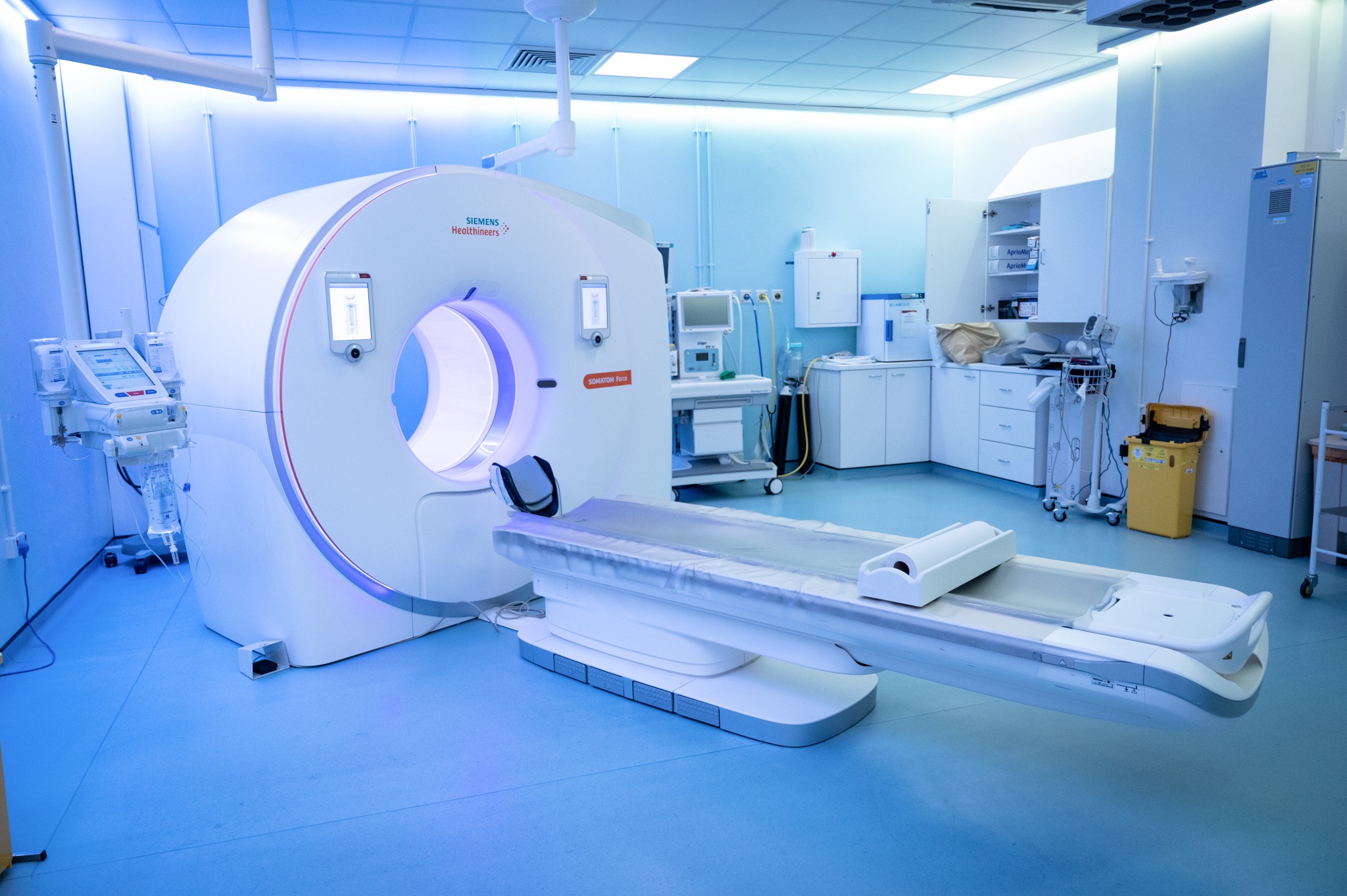
A conventional CT scanner has a short, ring-shaped opening and a motorised table. An X-ray source and detectors rotate around the area being examined, and a computer processes the measurements into cross-sectional images.
The radiologist reviews these images in different planes using display settings suited to the tissues being assessed. The machinery and positioning differ for specialist weight-bearing equipment.
CT does not use a strong magnetic field for imaging. Image acquisition is usually brief, but remaining still is important to reduce blurring. The radiographer can see and communicate with you throughout the examination.
Preparing for your ankle CT
Your centre will provide instructions. A routine non-contrast examination often needs little preparation. Contrast or specialist imaging may have additional requirements.
Before the appointment, tell the team about:
- Pregnancy or possible pregnancy.
- Kidney disease, dialysis or a recent change in kidney function.
- A previous reaction to CT contrast.
- Relevant medical conditions and regular medicines.
- Previous foot surgery, implants and earlier imaging.
- Pain, mobility needs or restrictions on standing or weight-bearing.
Metal near the scanned area can affect the images, so the team may ask you to remove footwear, jewellery or clothing containing metal. CT does not have the same magnetic restrictions as MRI.
Do not remove prescribed support or change weight-bearing restrictions without advice. Ask the centre about boots, casts or dressings beforehand. Bring relevant reports and any requested blood test results. Do not stop medicines or change existing fluid restrictions without clinical guidance.
What happens during the examination?
- Review and explanation. The team checks your referral, affected side, requested coverage and any contrast requirements.
- Preparing for imaging. You remove items near the foot that could affect image quality and may change clothing if required.
- Positioning. For a routine scan, the foot is supported on the scanner table. Specialist weight-bearing imaging uses separately planned positioning.
- Image acquisition. The table moves into position and images are obtained while you remain still and follow instructions.
- Additional images if needed. Contrast or further acquisitions may be used according to the protocol.
- Completion. The radiographer checks image quality and explains any observation or aftercare instructions.
Tell the team if positioning is painful or you need help moving. The radiographer obtains the images; a radiologist subsequently interprets them and prepares a written report.
Benefits, radiation and limitations
Ankle CT provides detailed bony information quickly and may support assessment of complex injuries or planning. A routine scan does not involve an incision.
CT uses ionising radiation. The expected clinical benefit and exposure are considered before scanning, and the protocol is selected to obtain useful information while managing exposure. Tell the team about pregnancy or possible pregnancy beforehand.
Contrast has separate suitability considerations and possible side effects, which are discussed if it is recommended. Image quality can also be affected by movement or metalwork.
CT does not identify the cause of every painful foot and may not be the best test for a tendon, ligament or early stress injury. Structural findings can be incidental, so the report needs clinical interpretation.
Appointment length and aftercare
A routine ankle CT appointment often takes around 15 to 30 minutes, with image acquisition taking only part of that time. Contrast, mobility assistance or specialist techniques may add time. Your centre will confirm how long to allow.
Most people can return to their usual routine after a standard examination. Continue any advice about a boot, crutches, weight-bearing or activity restrictions. Completing a scan does not establish that the foot is ready for unrestricted exercise.
If contrast is given, follow the centre’s observation and aftercare instructions, taking account of any existing fluid restrictions. The underlying foot problem may affect safe driving independently of the scan.
Getting and understanding your results
Your scan images are reviewed by a consultant radiologist or relevant specialist clinician, who prepares a written imaging report. Results are usually available within three working days of the date of your scan. More complex examinations may require additional specialist review and can take slightly longer.
Once your results are available, a post-scan consultation will be arranged with the same medical doctor who conducted your pre-scan consultation. This continuity allows your doctor to consider the findings alongside the symptoms, medical history and concerns you discussed before your scan.
During the consultation, your doctor will explain the report in detail, answer your questions and discuss what the findings mean for you. Where further assessment or care is needed, they will explain the recommended next steps, which may include additional investigations, a specialist referral or follow-up with your usual healthcare team.
How much does a private ankle CT cost?
PrivateScans CT scans start from £395. This is our general CT starting price. Your final cost depends on the chosen centre, required coverage and whether contrast or specialist imaging is needed.
Weight-bearing CT, assessment of both ankles or additional foot coverage may involve different arrangements and pricing. Our team will confirm the examination, price and included services before you proceed.
Arrange a private ankle CT
Your booking includes a 30-minute private telephone consultation with a medical doctor at a convenient time. The doctor will discuss your symptoms, injury history, previous investigations and treatment before advising whether ankle CT is clinically appropriate.
Where imaging is indicated, the referral identifies the affected side, region and clinical question. This helps the scanning team plan the protocol and gives the radiologist useful context.
You can explore our other CT body part examinations if a different area needs assessment. Urgent injuries and other concerning symptoms should be assessed promptly rather than waiting for a routine scan.
FAQs
What does an ankle CT show?
It provides detailed images of bones and joint surfaces within the requested coverage. It may help assess fractures, alignment, arthritis or a postoperative concern. Other tests may be needed for soft tissues.
Can CT diagnose every ankle sprain?
No. Ligament and tendon concerns may be better assessed clinically, with MRI or with ultrasound. Routine CT should not be assumed to exclude every soft tissue injury.
Does the scan include my whole foot?
Not automatically as a complete dedicated examination. Any additional foot coverage needs to be specified and arranged beforehand.
Will I stand for the scan?
Only if weight-bearing CT has been specifically requested and arranged. A routine examination is usually performed with the ankle supported on a scanner table.
Will I need a contrast injection?
Many routine examinations do not need contrast. Intravenous contrast or an arthrogram may be recommended for a particular question, and the team will explain any additional procedure.
How long does an ankle CT take?
Image acquisition is usually brief. Your centre will confirm the appointment length, including preparation, positioning and any additional imaging or observation.
When will my results be available?
Results are usually available within three working days of your scan. More complex examinations can take slightly longer. A post-scan consultation will be arranged with the same doctor who conducted your pre-scan consultation to explain the findings and any necessary next steps.
What’s included with your scan?
What is included with your scan:
- A pre-scan telephone consultation with a member of our medical team.
- A written medical referral prepared from the information discussed during your consultation, where clinically appropriate.
- Your scan at the chosen scanning centre.
- A written imaging report prepared by a consultant radiologist or relevant specialist clinician.
- Access to downloadable copies of your scan images where required.
- A post-scan consultation to discuss your results and next steps where needed.