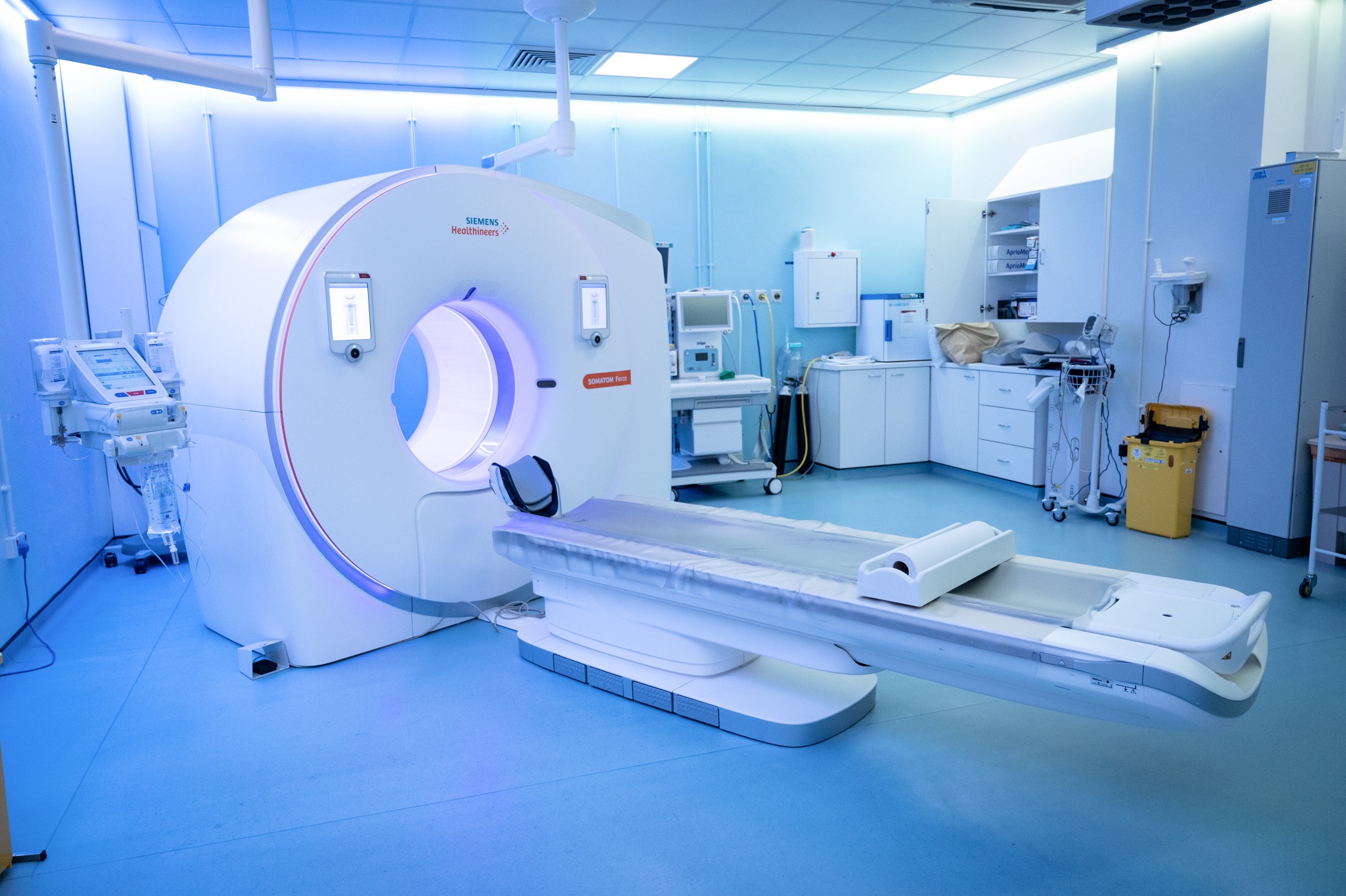
Scan overview
- Cardiac CT uses X-rays to produce detailed images of selected heart structures and blood vessels.
- Coronary CT angiography, calcium scoring and structural cardiac CT answer different clinical questions and use different protocols.
- A coronary CT angiogram uses intravenous contrast. Heart-rate medication and monitoring may also be needed, depending on the examination.
- Cardiac CT does not replace every cardiac investigation or emergency assessment for new or severe chest symptoms.
- PrivateScans CT scans start from £395. This is a general starting price; the team will confirm the price of your specialist cardiac examination.
What is a cardiac CT scan?
A cardiac CT is a computed tomography examination designed to assess the heart or selected associated blood vessels. It combines X-ray measurements taken from different angles to create detailed cross-sectional images that can also be reconstructed into additional views.
Because the heart moves continuously, cardiac protocols may coordinate image acquisition with the heartbeat using ECG monitoring. The required timing, coverage and use of contrast depend on the question being investigated.
The examination may assess the coronary arteries, selected heart structures or the anatomy needed to plan a procedure. A standard chest CT is not the same as a dedicated cardiac examination and does not automatically provide the same information.
Your clinician selects the appropriate investigation after considering symptoms, previous tests and relevant medical history. The images support clinical assessment rather than explaining every cause of chest discomfort, breathlessness or palpitations on their own.
Types of cardiac CT examination
Coronary CT angiography
A coronary CT angiogram, sometimes called CCTA or CTCA, uses an iodine-based contrast injection to assess the arteries supplying the heart muscle. It can help identify plaque and narrowing in the coronary arteries where this is the clinical concern.
Medication may be used to reduce heart rate or widen the coronary arteries, provided it is suitable for you. This is a diagnostic scan and does not involve placing a catheter in the coronary arteries to perform treatment.
Coronary calcium scoring
A calcium score examination measures calcified plaque in the coronary arteries using a non-contrast protocol. It may contribute to cardiovascular risk assessment in selected circumstances.
A calcium score is not the same as a coronary angiogram. It does not show every type of plaque or establish whether an artery is significantly narrowed. A low or zero score does not exclude every cause of chest symptoms.
Structural cardiac CT
Specialist protocols can assess selected anatomy involving the heart chambers, valves, aorta or related structures. They may be requested to plan a procedure or investigate a particular concern after treatment.
Not every cardiac booking includes all three examinations. The referral must identify the question and protocol required, and the centre must have the appropriate service available.
Why might cardiac CT be recommended?
Your clinician may recommend cardiac CT when the result is likely to clarify a cardiac question or influence management. The appropriate investigation depends on the symptoms and previous assessment.
Reasons may include:
- Assessment of suspected coronary artery disease after clinical review.
- Clarification of an uncertain result from another cardiac investigation.
- Selected cardiovascular risk assessment using an appropriate calcium score protocol.
- Assessment of a particular structural or congenital abnormality.
- Planning a valve or another specialist cardiac procedure.
- A defined question involving the aorta or changes after treatment.
Breathlessness, palpitations and chest symptoms can have several causes and may need other tests. Family history alone does not automatically determine that a coronary angiogram or calcium score is the appropriate next step.
New, severe or ongoing chest pain, marked breathlessness, collapse or other acute concerning symptoms needs urgent medical assessment. Do not wait for a routine private CT appointment.
What can cardiac CT help assess?
Coronary artery plaque and narrowing
A dedicated coronary angiogram can show selected plaque and assess narrowing in the arteries supplying the heart. The significance of a finding depends on its extent, image quality and the wider clinical picture. Further functional testing or invasive assessment may be needed in some cases.
Coronary calcium
A calcium score quantifies calcified coronary plaque. The result contributes to a broader risk discussion that includes age, medical history and other cardiovascular risk factors. It is not a direct measure of current blood flow or a standalone prediction of an individual event.
Congenital and structural differences
A tailored examination may define selected anatomical differences in the heart or vessels. It can complement echocardiography, cardiac MRI or another specialist investigation rather than replacing them in every case.
Valve and aortic anatomy
Specialist CT protocols can provide measurements and anatomical detail needed for selected valve procedures or assessment of the aorta. A routine coronary scan does not automatically include a complete investigation of all valve function or every aortic concern.
Pericardial and other tissue abnormalities
CT may help assess selected changes involving the pericardium, the tissue around the heart, or a particular mass. The diagnosis may still require other imaging or specialist tests. Imaging alone cannot definitively classify every abnormality.
Selected concerns after treatment
CT may be used for a defined question following surgery or another intervention. Previous reports and procedural details help the specialist interpret the findings and select the appropriate protocol.
Cardiac CT is not a universal test for heart failure, previous heart attack, rhythm disorders or every cardiac blood clot. These questions may require a different protocol or another investigation.
Cardiac CT and other heart investigations
A cardiac MRI provides different information about heart muscle, movement and selected tissue changes without ionising radiation. Echocardiography assesses cardiac structures and function using ultrasound, while ECG monitoring investigates electrical activity and rhythm.
An invasive coronary angiogram uses a catheter introduced through a blood vessel. It can provide additional assessment and, where appropriate, allow treatment such as stenting. A CT coronary angiogram is a diagnostic imaging examination and does not perform this treatment.
The most useful test depends on the clinical question. CT may be appropriate for one aspect of an investigation while another test is needed to assess symptoms or the functional importance of a finding.
When might another approach be needed?
A fast or irregular heartbeat can affect image quality. Heart-rate medication may help in some circumstances, but is not suitable for everyone and does not resolve every rhythm-related limitation.
Extensive coronary calcium, some stents or movement may make particular artery segments difficult to assess. A result may therefore lead to further investigation rather than a definitive answer from CT alone.
Reduced kidney function, a previous contrast reaction, pregnancy or other medical circumstances can affect the choice of protocol. None should be assessed from a general rule alone; the clinical and imaging teams consider your circumstances and the benefit of the proposed examination.
If another investigation is more appropriate, the team will explain the recommended approach.
Contrast and heart-rate medication
A coronary CT angiogram uses an iodine-based contrast injection through a cannula in a vein. A calcium score examination usually does not need contrast. Other structural protocols vary.
The team may give a beta blocker to reduce heart rate and a medicine such as glyceryl trinitrate, or GTN, to widen the coronary arteries. These medicines have their own suitability checks and possible side effects; they are not automatically given to every patient.
Tell the team about asthma or other breathing problems, low blood pressure, relevant heart conditions and all medicines you take. Medicines for erectile dysfunction, such as sildenafil, need particular discussion because they can interact with GTN. Follow the centre’s specific timing and medication instructions rather than altering treatment yourself.
Contrast suitability is assessed separately, including kidney function and previous contrast reactions. An injection can cause a temporary warm feeling or metallic taste. Report feeling unwell, breathing difficulty or other concerning symptoms immediately.
Preparing for cardiac CT
Your centre will provide instructions for your particular examination. These may include avoiding caffeine or nicotine for a specified period, fasting requirements and advice about regular medicines. Follow the exact instructions rather than assuming every cardiac CT has the same preparation.
Do not stop prescribed medicines or change fluid intake beyond existing restrictions without clinical advice. Contact the team if preparation could affect diabetes treatment or another medical condition.
Before the appointment, disclose:
- Kidney disease, dialysis or a recent change in kidney function.
- A previous reaction to CT contrast.
- Asthma, other breathing conditions, a slow or irregular heartbeat or low blood pressure.
- All medicines, including erectile dysfunction treatment.
- Pregnancy or possible pregnancy.
- Previous heart procedures, implants and relevant earlier investigations.
- Difficulty lying flat or holding your breath briefly.
Bring a medicine list, previous reports and kidney function results if requested. You may need to change into a gown so that ECG electrodes can be attached to the chest. Metal near the scanned area may need to be removed to avoid image distortion.
What happens during the examination?
- Review and checks. The team confirms the requested protocol, preparation, suitability and any contrast or medication requirements.
- Monitoring. ECG electrodes are placed on the chest to help coordinate scanning with the heartbeat. Heart rate and blood pressure may be checked.
- Medication if appropriate. Heart-rate treatment or GTN may be given according to the protocol, with time allowed for the intended effect.
- Positioning. You lie on the scanner table and the team helps you into the required position.
- Image acquisition. The table moves through the short scanner opening. Follow instructions about keeping still and holding your breath briefly.
- Contrast if required. An intravenous injection is given and images are timed to the appropriate phase.
- Completion and observation. The team checks the images and explains any monitoring or aftercare before you leave.
The radiographer and cardiac imaging team can see and communicate with you throughout. Tell them promptly if you are uncomfortable, feel unwell or cannot manage a breathing instruction.
Benefits, radiation and limitations
Cardiac CT can provide detailed anatomical information quickly without introducing a catheter into the coronary arteries. It may help clarify a diagnosis or plan a specialist procedure.
CT uses ionising radiation. The clinical benefit and exposure are considered before scanning, and the protocol is selected to obtain useful information while managing exposure. Tell the team about pregnancy or possible pregnancy beforehand.
Contrast and cardiac medicines have separate suitability considerations and potential side effects. Kidney function, previous reactions, blood pressure and relevant conditions are assessed according to the planned examination.
Image quality can be affected by rhythm, movement or extensive calcium. A normal or uncertain scan does not explain every symptom or replace all other heart investigations. Your clinician will discuss any limitations and whether further assessment is needed.
Appointment length and aftercare
Image acquisition is usually brief, but the appointment can take longer for monitoring, cannula placement, medication and observation. Your centre will confirm the time to allow for the specific protocol.
Many people return home the same day. Follow the team’s instructions about resuming activities and medicines, particularly if heart-rate treatment or GTN has been used. These medicines may cause temporary symptoms such as light-headedness or tiredness.
Ask about travelling home before the appointment and do not drive if you feel unwell or have been advised not to. Follow contrast aftercare instructions, taking account of any existing fluid restrictions or other medical advice.
Getting and understanding your results
Your scan images are reviewed by a consultant radiologist or relevant specialist clinician, who prepares a written imaging report. Results are usually available within three working days of the date of your scan. More complex examinations may require additional specialist review and can take slightly longer.
Once your results are available, a post-scan consultation will be arranged with the same medical doctor who conducted your pre-scan consultation. This continuity allows your doctor to consider the findings alongside the symptoms, medical history and concerns you discussed before your scan.
During the consultation, your doctor will explain the report in detail, answer your questions and discuss what the findings mean for you. Where further assessment or care is needed, they will explain the recommended next steps, which may include additional investigations, a specialist referral or follow-up with your usual healthcare team.
How much does a private cardiac CT cost?
CT coronary angiography starts from £1,095, and coronary artery calcium scoring starts from £445. These are starting prices; the examination and total cost are confirmed before booking.
The combined CTCA and calcium-scoring option starts from £1,540. Optional CaRi-Heart and CaRi-Plaque packages are explained on our Cardiac CT service page. Availability depends on the centre and clinical requirements.
Our team will confirm your selected examination, price and included services before you proceed.
Arrange a private cardiac CT
Your booking includes a 30-minute private telephone consultation with a medical doctor at a convenient time. The doctor will review your symptoms, relevant medical history, medicines and previous investigations before advising whether cardiac CT is clinically appropriate.
Where imaging is indicated, the clinical question and required specialist protocol are clarified so that the appropriate examination can be arranged. Your referral provides relevant context for the imaging team and reporting clinician.
You can explore our other CT body part examinations if a different area requires assessment. Acute chest symptoms need urgent clinical care rather than waiting for a routine scan.
FAQs
Is cardiac CT the same as a calcium score?
Calcium scoring is one type of cardiac CT. It measures calcified coronary plaque without contrast. A coronary CT angiogram uses contrast to assess the arteries in a different way, and the tests are not interchangeable.
Will I need contrast?
A coronary CT angiogram uses intravenous contrast, while calcium scoring usually does not. The team will confirm the protocol and assess suitability before the examination.
Why might I be given heart-rate medication?
A lower heart rate can improve coronary image quality in some protocols. Medication is given only where appropriate after relevant checks. Tell the team about all medicines and medical conditions beforehand.
Can I have the scan with an irregular heartbeat?
This needs individual assessment. Rhythm can affect image quality, and the team will decide whether the requested CT can provide useful information or another investigation is preferable.
Do I need to avoid caffeine?
Some protocols require this for a specified period. Follow your centre’s exact instructions about caffeine, nicotine, food and medicines.
Can I drive home afterwards?
Ask the team in advance, particularly if medication is planned. Do not drive if you feel light-headed or unwell or have been advised to arrange a lift. Follow the specific aftercare instructions.
When will my results be available?
Results are usually available within three working days of your scan. More complex examinations can take slightly longer. A post-scan consultation will be arranged with the same doctor who conducted your pre-scan consultation to explain the findings and any necessary next steps.
What’s included with your scan?
What is included with your scan:
- A pre-scan telephone consultation with a member of our medical team.
- A written medical referral prepared from the information discussed during your consultation, where clinically appropriate.
- Your scan at the chosen scanning centre.
- A written imaging report prepared by a consultant radiologist or relevant specialist clinician.
- Access to downloadable copies of your scan images where required.
- A post-scan consultation to discuss your results and next steps where needed.